| CHAPTER VI.
MINOR TERATA. Anomalies and Curiosities of Medicine | ||
6. CHAPTER VI.
MINOR TERATA.
Ancient Ideas Relative to Minor Terata.—The ancients viewed with great interest the minor structural anomalies of man, and held them to be divine signs or warnings in much the same manner as they considered more pronounced monstrosities. In a most interesting and instructive article, Ballantyne [6.1] quotes Ragozin in saying that the Chaldeo-Babylonians, in addition to their other numerous subdivisions of divination, drew presages and omens for good or evil from the appearance of the liver, bowels, and viscera of animals offered for sacrifice and opened for inspection, and from the natural defects or monstrosities of babies or the young of animals. Ballantyne names this latter subdivision of divination fetomancy or teratoscopy, and thus renders a special chapter as to omens derived from monstrous births, given by Lenormant:—
"The prognostics which the Chaldeans claimed to draw from monstrous births in man and the animals are worthy of forming a class by themselves, insomuch the more as it is the part of their divinatory science with which, up to the present time, we are best acquainted. The development that their astrology had given to `généthliaque,' or the art of horoscopes of births, had led them early to attribute great importance to all the teratologic facts which were there produced. They claimed that an experience of 470,000 years of observations, all concordant, fully justified their system, and that in nothing was the influence of the stars marked in a more indubitable manner than in the fatal law which determined the destiny of each individual according to the state of the sky at the moment when he came into the world. Cicero, by the very terms which he uses to refute the Chaldeans, shows that the result of these ideas was to consider all infirmities and monstrosities that new-born infants exhibited as the inevitable and irremediable consequence of the action of these astral positions. This being granted, the observation of similar monstrosities gave, as it were, a reflection of the state of the sky; on which depended all terrestrial things; consequently, one might read in them the future with as much certainty as in the stars themselves. For this reason the greatest possible importance was attached to the teratologic auguries which occupy so much space in the fragments of the great
The rendering into English of the account of 62 teratologic cases in the human subject with the prophetic meanings attached to them by Chaldean diviners, after the translation of Opport, is given as follows by Ballantyne, some of the words being untranslatable:—
"When a woman gives birth to an infant—
(1) that has the ears of a lion, there will be a powerful king in the country;
(2) that wants the right ear, the days of the master (king) will be prolonged (reach old age);
(3) that wants both ears, there will be mourning in the country, and the country will be lessened (diminished);
(4) whose right ear is small, the house of the man (in whose house the birth took place) will be destroyed;
(5) whose ears are both small, the house of the man will be built of bricks;
(6) whose right ear is mudissu tehaat (monstrous), there will be an androgyne in the house of the new-born
(7) whose ears are both mudissu (deformed), the country will perish and the enemy rejoice;
(8) whose right ear is round, there will be an androgyne in the house of the new-born;
(9) whose right ear has a wound below, and tur re ut of the man, the house will be destroyed;
(10) that has two ears on the right side and none on the left, the gods will bring about a stable reign, the country will flourish, and it will be a land of repose;
(11) whose ears are both closed, sa a au;
(12) that has a bird's beak, the country will be peaceful;
(13) that has no mouth, the mistress of the house will die;
(14) that has no right nostril, the people of the world will be injured;
(15) whose nostrils are absent, the country will be in affliction, and the house of the man will be ruined;
(16) whose jaws are absent, the days of the master (king) will be prolonged, but the house (where the infant is born) will be ruined.
When a woman gives birth to an infant—
(17) that has no lower jaw, mut ta at mat, the name will not be effaced;
(20) that has no nose, affliction will seize upon the country, and the master of the house will die;
(21) that has neither nose nor virile member (penis), the army of the king will be strong, peace will be in the land, the men of the king will be sheltered from evil influences, and Lilit (a female demon) shall not have power over them;
(22) whose upper lip overrides the lower, the people of the world will rejoice (or good augury for the troops);
(23) that has no lips, affliction will seize upon the land, and the house of the man will be destroyed;
(24) whose tongue is kuri aat, the man will be spared (?);
(25) that has no right hand, the country will be convulsed by an earthquake;
(26) that has no fingers, the town will have no births, the barshall be lost;
(27) that has no fingers on the right side, the master (king) will not pardon his adversary (or shall be humiliated by his enemies);
(28) that has six fingers on the right side, the man will take the lukunu of the house;
(29) that has six very small toes on both feet, he shall not go to the lukunu;
(30) that has six toes on each foot, the people of the world will be injured (calamity to the troops);
(32) that has no penis, the master of the house will be enriched by the harvest of his field;
(33) that wants the penis and the umbilicus, there will be ill-will in the house, the woman (wife) will have an overbearing eye (be haughty); but the male descent of the palace will be more extended.
When a woman gives birth to an infant—
(34) that has no well-marked sex, calamity and affliction will seize upon the land; the master of the house shall have no happiness;
(35) whose anus is closed, the country will suffer from want of nourishment;
(36) whose right testicle (?) is absent, the country of the master (king) will perish;
(37) whose right foot is absent, his house will be ruined and there will be abundance in that of the neighbor;
(38) that has no feet, the canals of the country will be cut (intercepted) and the house ruined;
(39) that has the right foot in the form of a fish's tail, the booty of the country of the humble will not be imas sa bir;
(40) whose hands and feet are like four fishes' tails (fins), the master (king) shall perish (?) and his country shall be consumed;
(41) whose feet are moved by his great hunger, the house of the su su shall be destroyed;
(42) whose foot hangs to the tendons of the body, there will be great prosperity in the land;
(43) that has three feet, two in their normal position (attached to the body) and the third between them, there will be great prosperity in the land;
(44) whose legs are male and female, there will be rebellion;
(45) that wants the right heel, the country of the master (king) will be destroyed.
When a woman gives birth to au infant—
(46) that has many white hairs on the head, the days of the king will be prolonged;
(47) that has much ipga on the head, the master of the house will die, the house will be destroyed;
(48) that has much pinde on the head, joy shall go to meet the house (that has a head on the head, the good augury shall enter at its aspect into the house);
(49) that has the head full of hali, there will be ill-will toward him and the master (king) of the town shall die;
(50) that has the head full of siksi the king will repudiate his masters;
(51) that has some pieces of flesh (skin) hanging on the head, there shall be ill-will;
(52) that has some branches (?) (excrescences) of flesh (skin) hanging on the head, there shall be ill-will, the house will perish;
(53) that has some formed fingers (horns ?) on the head, the days of the king will be less and the years lengthened (in the duration of his old age);
(54) that has some kali on the head, there will be a king of the land;
(55) that has a — of a bird on the head, the master of the house shall not prosper;
(56) that has some teeth already through (cut), the days of the king will arrive at old age, the country will show itself powerful over (against) strange (feeble) lands, but the house where the infant is born will be ruined;
(57) that has the beard come out, there will be abundant rains;
(58) that has some birta on the head, the country will be strengthened (reinforced);
(59) that has on the head the mouth of an old man and that foams (slabbers), there will be great prosperity in the land, the god Bin will give a magnificent harvest (inundate the land with fertility), and abundance shall be in the land;
(60) that has on one side of the head a thickened ear, the first-born of the men shall live a long time (?);
(62) that has the figure in horn (like a horn ?) . . .''
As ancient and as obscure as are these records, Ballantyne has carefully gone over each, and gives the following lucid explanatory comments:—
"What `ears like a lion' (No. 1) may have been it is difficult to determine; but doubtless the direction and shape of the auricles were so altered as to give them an animal appearance, and possibly the deformity was that called `orechio ad ansa' by Lombroso. The absence of one or both ears (Nos. 2 and 3) has been noted in recent times by Virchow (Archiv für path. Anat.. xxx., p. 221), Gradenigo (Taruffi's `Storia della Teratologia,' vi., p. 552), and others. Generally some cartilaginous remnant is found, but on this point the Chaldean record is silent. Variations in the size of the ears (Nos. 4 and 5) are well known at the present time, and have been discussed at length by Binder (Archiv für Psychiatrie und Nervenkrankheiten, xx., 1887) and others. The exact malformation indicated in Nos. 6 and 7 is, of course, not to be determined, although further researches in Assyriology may clear up this point. The `round ear' (No. 8) is one of Binder's types, and that with a `wound below' (No. 9) probably refers to a case of fistula auris congenita (Toynbee, `Diseases of the Ear,' 1860). The instance of an infant born with two ears on the right side (No. 10) was doubtless one of cervical auricle or preauricular appendage, whilst closure of the external auditory meatus (No. 11) is a well-known deformity.
"The next thirteen cases (Nos. 12-24) were instances of anomalies of the mouth and nose. The `bird's beak' (No. 12) may have been a markedly aquiline nose; No. 13 was a case of astoma; and Nos. 14 and 15 were instances of stenosis or atresia of the anterior nares. Fetuses with absence of the maxillæ (Nos. 16 and 17) are in modern terminology called agnathous. Deformities like that existing in Nos. 20 and 21 have been observed in paracephalic and cyclopic fetuses. The coincident absence of nose and penis (No. 21) is interesting, especially when taken in conjunction with the popular belief that the size of the former organ varies with that of the latter. Enlargement of the upper lip (No. 22), called epimacrochelia by Taruffi, and absence of the lips (No. 23), known now under the name of brachychelia, have been not unfrequently noticed in recent times. The next six cases (Nos. 25-30) were instances of malformations of the upper limb: Nos. 25, 26. and 27 were probably instances of the so-called spontaneous or intrauterine amputation; and Nos. 28, 29, and 30 were examples of the comparatively common deformity known as polydactyly. No. 31 was probably a case of ectopia cordis.
"Then follow five instances of genital abnormalities ( Nos. 32-36), consisting of absence of the penis (epispadias?), absence of penis and umbilicus (epispadias and exomphalos?), hermaphroditism, imperforate anus, and nondescent of one testicle. The nine following cases (Nos. 37-45) were anomalies of the lower limbs: Nos. 37, 38, and 42 may have been spontaneous amputations; Nos. 39 and 40 were doubtless instances of webbed toes (syndactyly), and the deformity indicated in No. 45 was presumably talipes equinus. The infant born with three feet (No. 43) was possibly a case of parasitic monstrosity, several of which have been reported in recent teratologic literature; but what is meant by the statement concerning `male and female legs' it is not easy to determine.
"Certain of the ten following prodigies (Nos. 46-55) cannot in the present state of our knowledge be identified. The presence of congenital patches of white or gray hair on the scalp, as recorded in No. 46, is not an unknown occurrence at the present time; but what the Chaldeans meant by ipga, pinde, hali riksi, and kali on the head of the new-born infant it is impossible to tell. The guess may be hazarded that cephalhematoma, hydrocephalus, meningocele, nevi, or an excessive amount of vernix caseosa were the conditions indicated, but a wider acquaintance with the meaning of the cuneiform characters is necessary before any certain identification is possible. The `pieces of skin hanging from the
"The remaining observations (No. 56-62) refer to cases of congenital teeth (No. 56) to deformity of the ears (Nos. 60 and 61), and a horn (No. 62).''
From these early times almost to the present day similar significance has been attached to minor structural anomalies. In the following pages the individual anomalies will be discussed separately and the most interesting examples of each will be cited. It is manifestly evident that the object of this chapter is to mention the most striking instances of abnormism and to give accompanying descriptions of associate points of interest, rather than to offer a scientific exposition of teratology, for which the reader is referred elsewhere.
Congenital defect of the epidermis and true skin is a rarity in pathology. Pastorello [6.2] speaks of a child which lived for two and a half hours whose hands and feet were entirely destitute of epidermis; the true skin of those parts looked like that of a dead and already putrefying child. Hanks [6.3] cites the history of a case of antepartum desquamation of the skin in a living fetus. Hochstetter [6.4] describes a full-term, living male fetus with cutaneous defect on both sides of the abdomen a little above the umbilicus. The placenta and membranes were normal, a fact indicating that the defect was not due to amniotic adhesions; the child had a club-foot on the left side. The mother had a fall three weeks before labor.
Abnormal Elasticity of the Skin.—In some instances the skin is affixed so loosely to the underlying tissues and is possessed of so great elasticity that it can be stretched almost to the same extent as India rubber. There have been individuals who could take the skin of the forehead and pull it down over the nose, or raise the skin of the neck over the mouth. They also occasionally have an associate muscular development in the subcutaneous tissues similar to the panniculus adiposus of quadrupeds, giving them preternatural motile power over the skin. The man recently exhibited under the title of the "Elastic-Skin Man'' was an example of this anomaly. The first of this class of exhibitionists was seen in Buda-Pesth some years since and possessed great elasticity in the skin of his whole body; even his nose could be stretched. Figure 70 represents a photograph of an exhibitionist named Felix Wehrle, who besides having the power to stretch his skin could readily bend his fingers backward and forward. The photograph was taken in January, 1888.
In these congenital cases there is loose attachment of the skin without hypertrophy, to which the term dermatolysis is restricted by Crocker. Job van Meekren, *[560] the celebrated Dutch physician of the seventeenth century,
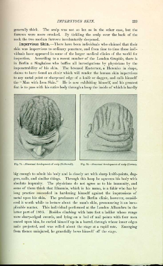
Occasionally abnormal development of the scalp is noticed. McDowall [6.5] of twenty-two. On each side of the median line of the head there were five deep furrows (Fig. 71), more curved and shorter as the distance from the median line increased. In the illustration the hair in the furrows is left longer than that on the rest of the head. The patient was distinctly microcephalic and the right side of the body was markedly wasted. The folds were due to hypertrophy of the muscles and scalp, and the
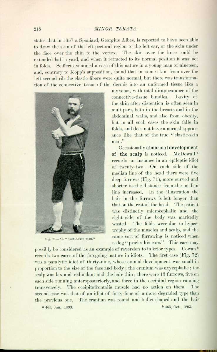
Fig. 70.—An "elastic-skin man.''
[Description: Photograph of a man stretching the skin of his arm]
same sort of furrowing is noticed when a dog "pricks his ears.'' This case may possibly be considered as an example of reversion to inferior types. Cowan [6.6] records two cases of the foregoing nature in idiots. The first case (Fig. 72) was a paralytic idiot of thirty-nine, whose cranial development was small in proportion to the size of the face and body; the cranium was oxycephalic; the scalp was lax and redundant and the hair thin; there were 13 furrows, five on each side running anteroposteriorly, and three in the occipital region running transversely. The occipitofrontalis muscle had no action on them. The second case was that of an idiot of forty-four of a more degraded type than the previous one. The cranium was round and bullet-shaped and the hair
Impervious Skin.—There have been individuals who claimed that their skin was impervious to ordinary puncture, and from time to time these individuals have appeared in some of the larger medical clinics of the world for inspection. According to a recent number of the London Graphic, there is in Berlin a Singhalese who baffles all investigations by physicians by the impenetrability of his skin. The bronzed Easterner, a Hercules in shape, claims to have found an elixir which will render the human skin impervious to any metal point or sharpened edge of a knife or dagger, and calls himself the "Man with Iron Skin.'' He is now exhibiting himself, and his greatest feat is to pass with his entire body through a hoop the inside of which is hardly

big enough to admit his body and is closely set with sharp knife-points, daggers, nails, and similar things. Through this hoop he squeezes his body with absolute impunity. The physicians do not agree as to his immunity, and some of them think that Rhannin, which is his name, is a fakir who has by long practice succeeded in hardening himself against the impressions of metal upon his skin. The professors of the Berlin clinic, however, considered it worth while to lecture about the man's skin, pronouncing it an inexplicable matter. This individual performed at the London Alhambra in the latter part of 1895. Besides climbing with bare feet a ladder whose rungs were sharp-edged swords, and lying on a bed of nail points with four men seated upon him, he curled himself up in a barrel, through whose inner edges nails projected, and was rolled about the stage at a rapid rate. Emerging from thence uninjured, he gracefully bows himself off the stage.
Albinism is characterized by the absolute or relative absence of pigment of the skin, due to an arrest, insufficiency, or retardation of this pigment. Following Trélat and Guinard, we may divide albinism into two classes,—general and partial.
As to the etiology of albinism, there is no known cause of the complete form. Heredity plays no part in the number of cases investigated by the authors. D'Aubé, by his observations on white rabbits, believes that the influence of consanguinity is a marked factor in the production of albinism; there are, however, many instances of heredity in this anomaly on record, and this idea is possibly in harmony with the majority of observers. Geoffroy-Saint-Hilaire has noted that albinism can also be a consequence of a pathologic condition
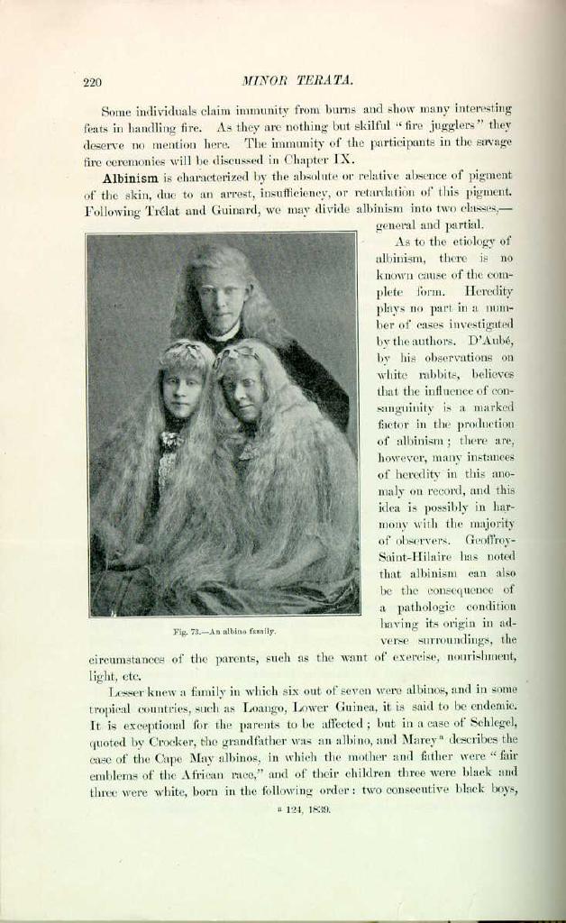
Fig. 73.—An albino family.
[Description: Photograph of three members of an albino family]
having its origin in adverse surroundings, the circumstances of the parents, such as the want of exercise, nourishment, light, etc.
Lesser knew a family in which six out of seven were albinos, and in some tropical countries, such as Loango, Lower Guinea, it is said to be endemic. It is exceptional for the parents to be affected; but in a case of Schlegel, quoted by Crocker, the grandfather was an albino, and Marey [6.7] describes the case of the Cape May albinos, in which the mother and father were "fair emblems of the African race,'' and of their children three were black and three were white, born in the following order: two consecutive black boys,
Examples of the total absence of pigment occur in all races, but particularly is it interesting when seen in negroes who are found absolutely white but preserving all the characteristics of their race, as, for instance, the kinky, woolly hair, flattened nose, thick lips, etc. René Claillé, in his "Voyage à Tombouctou,'' says that he saw a white infant, the offspring of a negro and negress. Its hair was white, its eyes blue, and its lashes flaxen. Its pupils were of a reddish color, and its physiognomy that of a Mandingo. He says such cases are not at all uncommon; they are really negro albinos. Thomas Jefferson, in his "History of Virginia,'' has an excellent description of these negroes, with their tremulous and weak eyes; he remarks that they freckle easily. Buffon speaks of Ethiops with white twins, and says that albinos are quite common in Africa, being generally of delicate constitution, twinkling eyes, and of a low degree of intelligence; they are despised and ill-treated by the other negroes. Prichard, quoted by Sedgwick, speaks of a case of atavic transmission of albinism through the male line of the negro race. The grandfather and the grandchild were albinos, the father being black. There is a case [6.9] of a brother and sister who were albinos, the parents being of ordinary color but the grandfather an albino. Coinde, quoted by Sedgwick, speaks of a man who, by two different wives, had three albino children.
A description of the ordinary type of albino would be as follows: The skin and hair are deprived of pigment; the eyebrows and eyelashes are of a brilliant white or are yellowish; the iris and the choroid are nearly or entirely deprived of coloring material, and in looking at the eye we see a roseate zone and the ordinary pink pupil; from absence of pigment they necessarily keep their eyes three-quarters closed, being photophobic to a high degree. They are amblyopic, and this is due partially to a high degree of ametropia (caused by crushing of the eyeball in the endeavor to shut out light) and from retinal exhaustion and nystagmus. Many authors have claimed that they have little intelligence, but this opinion is not true. Ordinarily the reproductive functions are normal, and if we exclude the results of the union of two albinos we may say that these individuals are fecund.
Partial albinism is seen. The parts most often affected are the genitals, the hair, the face, the top of the trunk, the nipple, the back of the hands and fingers. Folker [6.10] reports the history of a case of an albino girl having pink eyes and red hair, the rest of the family having pink eyes and white
Albinism is found in the lower animals, and is exemplified ordinarily by rats, mice, crows, robins, etc. In the Zoologic Garden at Baltimore two years ago was a pair of pure albino opossums. The white elephant is celebrated in the religious history of Oriental nations, and is an object of veneration and worship in Siam. White monkeys and white roosters are also worshiped. In the Natural History Museum in London there are stuffed examples of albinism and melanism in the lower animals.
Melanism is an anomaly, the exact contrary of the preceding. It is characterized by the presence in the tissues and skin of an excessive amount of pigment. True total melanism is unknown in man, in whom is only observed partial melanism, characterized simply by a pronounced coloration of part of the integument.
Some curious instances have been related [6.11] of an infant with a two-colored face, and of others with one side of the face white and the other black; whether they were cases of partial albinism or partial melanism cannot be ascertained from the descriptions.
Such epidermic anomalies as ichthyosis, scleroderma, and molluscum simplex, sometimes appearing shortly after birth, but generally seen later in life, will be spoken of in the chapter on Anomalous Skin Diseases.
Human horns are anomalous outgrowths from the skin and are far more frequent than ordinarily supposed. Nearly all the older writers cite examples. Aldrovandus, Amatus Lusitanus, Boerhaave, Dupré, Schenck, Riverius, Vallisneri, and many others mention horns on the head. In the ancient times horns were symbolic of wisdom and power. Michael Angelo in his famous sculpture of Moses has given the patriarch a pair of horns. Rhodius *[680] observed a Benedictine monk who had a pair of horns and who was addicted to rumination. Fabricius *[333] saw a man with horns on his head, whose son ruminated; the son considered that by virtue of his ruminating characteristics his father had transmitted to him the peculiar anomaly of the family. Fabricius Hildanus *[334] saw a patient with horns all over the body and another with horns on the forehead. Gastaher [6.12] speaks of a horn from the left temple; Zacutus Lusitanus *[831] saw a horn from the heel; Wroe, *[629] one of considerable length from the scapula; Cosnard, one from the bregma; the Ephemerides, from the foot; Borellus, from the face and foot, and Ash, [6.13] horns all over the body. Home, Cooper, and Treves have collected examples of horns, and there is one 11 inches long and 2 1/2 in circumference in a London museum. Lozes collected reports of 71 cases of horns,—37 in females, 31 in males, and three in infants. Of this number, 15 were on the head, eight on the face, 18 on the lower extremities, eight on the trunk, and three on the glans
Instances of cutaneous horns, when seen and reported by the laity, give rise to most amusing exaggerations and descriptions. The following account [6.16] is given in New South Wales, obviously embellished with apocryphal details by some facetious journalist: The child, five weeks old, was born with hair two inches long all over the body; his features were fiendish and his eyes shone like beads beneath his shaggy brows. He had a tail 18 inches long, horns from the skull, a full set of teeth, and claw-like hands; he snapped like a dog and crawled on all fours, and refused the natural sustenance of a normal child. The mother almost became an imbecile after the birth of the monster. The country people about Bomballa considered this devil-child a punishment for a rebuff that the mother gave to a Jewish peddler selling Crucifixion-pictures. Vexed by his persistence, she said she would sooner have a devil in her house than his picture.
Lamprey [6.17] has made a minute examination of the much-spoken-of "Horned Men of Africa.'' He found that this anomaly was caused by a congenital malformation and remarkable development of the infraorbital ridge of the maxillary bone (Fig. 74). He described several cases, and through an interpreter found that they were congenital, followed no history of traumatism, caused little inconvenience, and were unassociated with disturbance of the sense of smell. He also learned that the deformity was quite rare in the Cape Coast region, and received no information tending to prove the conjecture that the tribes in West Africa used artificial means to produce the anomaly, although such custom is prevalent among many aborigines.
Probably the most remarkable case of a horn was that of Paul Rodrigues, a Mexican porter, [6.18] who, from the upper and lateral part of his head, had a horn 14 inches in circumference and divided into three shafts, which he concealed by constantly wearing a peculiarly shaped red cap. There is in Paris a wax model of a horn, eight or nine inches in length, removed from an old woman by the celebrated Souberbielle. Figure 75 is from a wax model supposed to have been taken from life, showing an enormous grayish-black horn proceeding from the forehead. Warren mentions a case under the care of Dubois, in a woman from whose forehead grew a horn six inches in diameter and six inches in height. It was hard at the summit and had a fetid
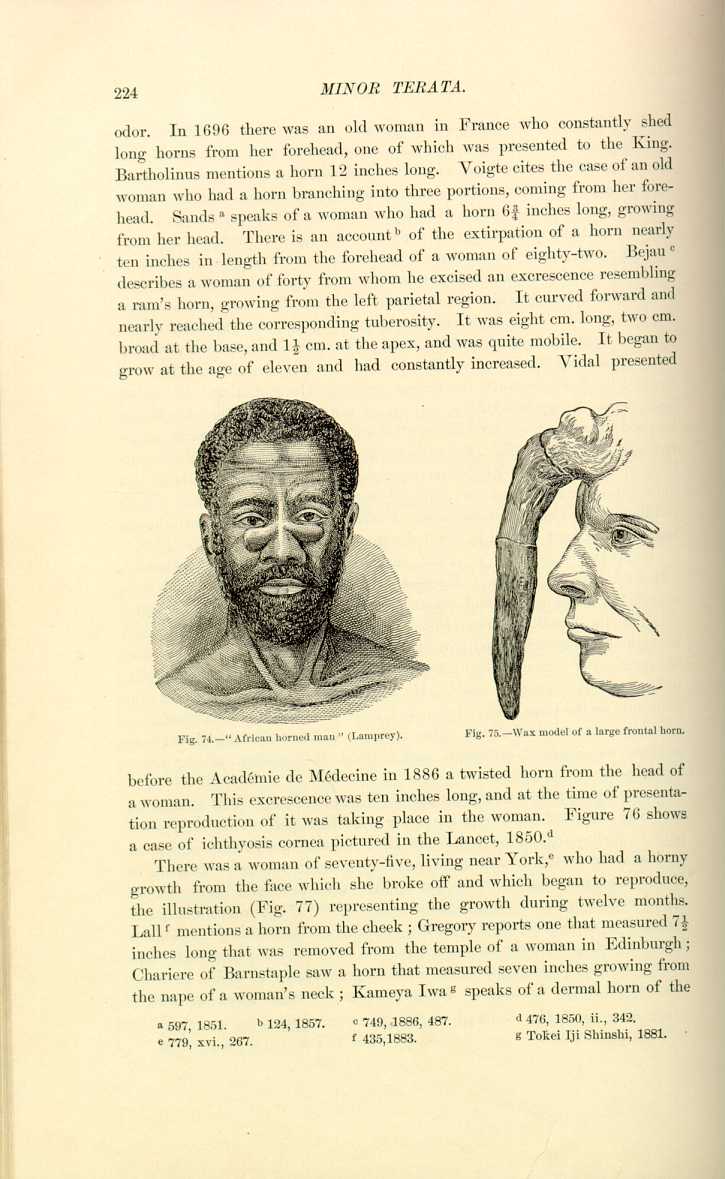
Fig. 74.—"African horned man'' (Lamprey).
[Description: Drawing of an African horned man]

Fig. 75.—Wax model of a large frontal horn.
[Description: Drawing of a wax model of a frontal horn]
before the Académie de Médecine in 1886 a twisted horn from the head of a woman. This excrescence was ten inches long, and at the time of presentation reproduction of it was taking place in the woman. Figure 76 shows a case of ichthyosis cornea pictured in the Lancet, 1850. [6.22]
There was a woman of seventy-five, living near York, [6.23] who had a horny growth from the face which she broke off and which began to reproduce, the illustration (Fig. 77) representing the growth during twelve months. Lall [6.24] mentions a horn from the cheek; Gregory reports one that measured 7 1/2 inches long that was removed from the temple of a woman in Edinburgh; Chariere of Barnstaple saw a horn that measured seven inches growing from the nape of a woman's neck; Kameya Iwa [6.25] speaks of a dermal horn of the

Fig. 76.—Ichthyosis cornea.
[Description: Drawing of an ichtyosis cornea]
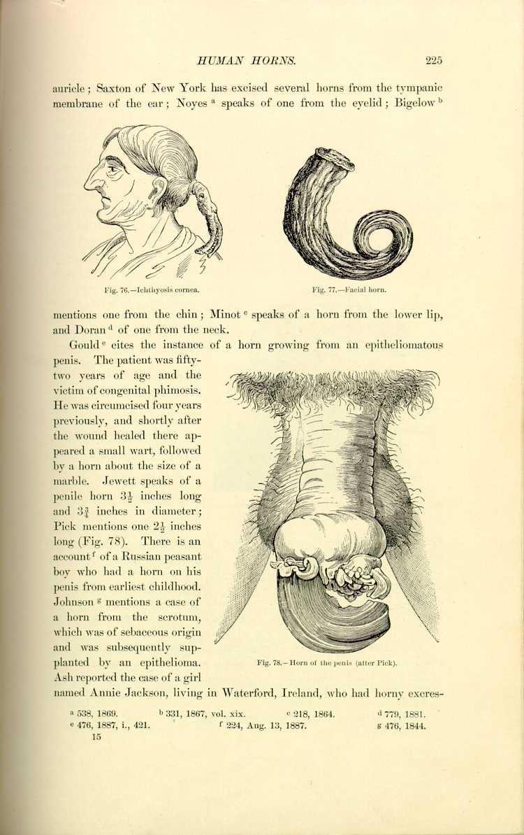
Fig. 77.—Facial horn.
[Description: Drawing of a facial horn]
mentions one from the chin; Minot [6.28] speaks of a horn from the lower lip, and Doran [6.29] of one from the neck.
Gould [6.30] cites the instance of a horn growing from an epitheliomatous penis. The patient was fifty-two years of age and the victim of congenital phimosis. He was circumcised four years previously, and shortly after the wound healed there appeared a small wart, followed by a horn about the size of a marble. Jewett speaks of a penile horn 3 1/2 inches long and 3 3/4 inches in diameter; Pick mentions one 2 1/2 inches long (Fig. 78). There is an account [6.31] of a Russian peasant boy who had a horn on his penis from his earliest childhood. Johnson [6.32] mentions a case of a horn from the scrotum, which was of sebaceous origin and was subsequently supplanted by an epithelioma.

Fig. 78.—Horn of the penis (after Pick).
[Description: Drawing of a penis with a horn]
Ash reported the case of a girl named Annie Jackson, living in Waterford, Ireland, who had horny excrescences
Wagstaffe [6.33] presents a horn which grew from the middle of the leg six inches below the knee in a woman of eighty. It was a flattened spiral of more than two turns, and during forty years' growth had reached the length of 14.3 inches. Its height was 3.8 inches, its skin-attachment 1.5 inches in diameter, and it ended in a blunt extremity of 0.5 inch in diameter. Stephens [6.34] mentions a dermal horn on the buttocks at the seat of a carcinomatous cicatrix. Harris [6.35] and Domonceau [6.36] speak of horns from the leg. Cruveilhier [6.37] saw a Mexican Indian who had a horn four inches long and eight inches in circumference growing from the left lumbar region. It had been sawed off twice by the patient's son and was finally extirpated by Faget. The length of the pieces was 12 inches. Bellamy [6.38] saw a horn on the clitoris about the size of a tiger's claw in a its origin from beneath the preputium clitoridis.
Horns are generally solitary but cases of multiple formation are known Lewin and Heller record a syphilitic case with eight cutaneous horns on the palms and soles. A female patient of Manzuroff had as many as 185 horns.
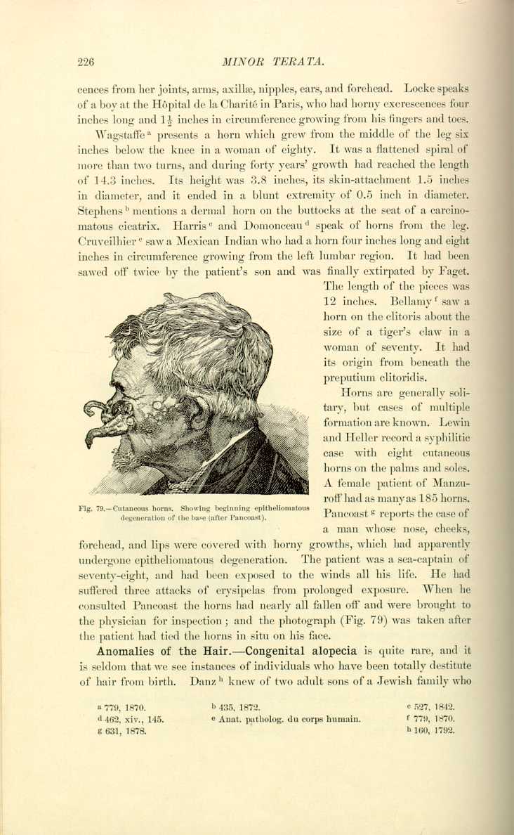
Pancoast [6.39] reports the case of a man whose nose, cheeks, forehead, and lips were covered with horny growths, which had apparently undergone epitheliomatous degeneration. The patient was a sea-captain of seventy-eight, and had been exposed to the winds all his life. He had suffered three attacks of erysipelas from prolonged exposure. When he consulted Pancoast the horns had nearly all fallen off and were brought to the physician for inspection; and the photograph (Fig. 79) was taken after the patient had tied the horns in situ on his face.
Anomalies of the Hair.—Congenital alopecia is quite rare, and it is seldom that we see instances of individuals who have been totally destitute of hair from birth. Danz [6.40] knew of two adult sons of a Jewish family who
Hutchinson [6.44] mentions a boy of three and a half in whom there was congenital absence of hair and an atrophic condition of the skin and appendages. His mother was bald from the age of six, after alopecia areata. Schede reports two cases of congenitally bald children of a peasant woman (a boy of thirteen and a girl of six months). They had both been born quite bald, and had remained so. In addition there were neither eyebrows nor eyelashes and nowhere a trace of lanugo. The children were otherwise healthy and well formed. The parents and brothers were healthy and possessed a full growth of hair. Thurman [6.45] reports a case of a man of fifty-eight, who was almost devoid of hair all his life and possessed only four teeth. His skin was very delicate and there was absence of sensible perspiration and tears. The skin was peculiar in thinness, softness, and absence of pigmentation. The hair on the crown of the head and back was very fine, short, and soft, and not more in quantity than that of an infant of three months. There was a similar peculiarity in his cousin-german. Williams mentions the case of a young lady of fifteen with scarcely any hair on the eyebrows or head and no eyelashes. She was edentulous and had never sensibly perspired. She improved under tonic treatment.
Instances are on record of women devoid of hair about the genital region. Riolan says that he examined the body of a female libertine who was totally hairless from the umbilical region down.
Congenital alopecia is seen in animals. There is a species of dog, a native of China but now bred in Mexico and in the United States, which is distinguished for its congenital alopecia. The same fact has been observed occasionally in horses, cattle, and dogs. Heusner [6.46] has seen a pigeon destitute of feathers, and which engendered a female which in her turn transmitted the same characteristic to two of her young.
Sexualism and Hair Growth.—The growth or development of the hair may be accelerated by the state of the organs of generation. This is peculiarly noticeable in the pubic hairs and the beard, and is fully exemplified in the section on precocious development (Chapter VII.); however, Moreau de la Sarthe showed a child to the Medical Faculty of Paris in whom precocious development of the testicles had influenced that of the hair to such a degree that, at the age of six, the chest of this boy was as thickly set with hair as is usually seen in adults. It is well known that eunuchs often lose a great part of their beards, and after removal of the ovaries women are seen to develop an extra quantity of hair. Gerberon [6.47] tells of an infant with a beard, and Paullini [6.48] and the Ephemerides mention similar instances.
Bearded women are not at all infrequent. Hippocrates mentions a female who grew a beard shortly after menstruation had ceased. It is a well-recognized fact that after the menopause women become more hirsute, the same being the case after removal of any of the functional generative apparatus. Vicat saw a virgin who had a beard, and Joch [6.49]
Barbara Urster, who lived in the 16th century, had a beard to her girdle. The most celebrated "bearded woman'' was Rosine-Marguerite Müller, who died in a hospital in Dresden in 1732, with a thick beard and heavy mustache. Julia Pastrana had her face covered with thick hair and had a full beard and mustache. She exhibited defective dentition in both jaws, and the

Fig. 80.—Julia Pastrana.
[Description: Drawing of Julia Pastrana]
teeth present were arranged in an irregular fashion. She had pronounced prognathism, which gave her a simian appearance (Fig. 80). Ecker examined in 1876 a woman who died at Fribourg, whose face contained a full beard and a luxuriant mustache.
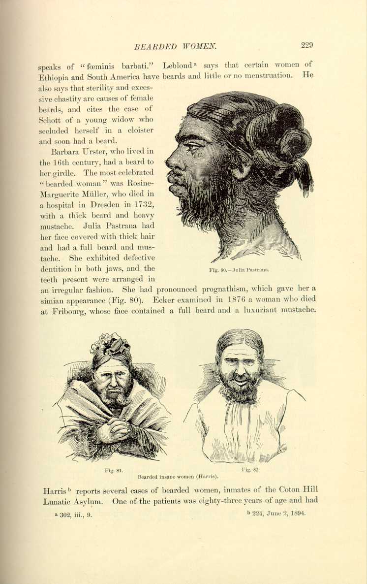
Fig. 81. Bearded insane women (Harris). Fig. 82.
[Description: Drawing of bearded women]
Harris [6.51] reports several cases of bearded women, inmates of the Coton Hill Lunatic Asylum. One of the patients was eighty-three years of age and had
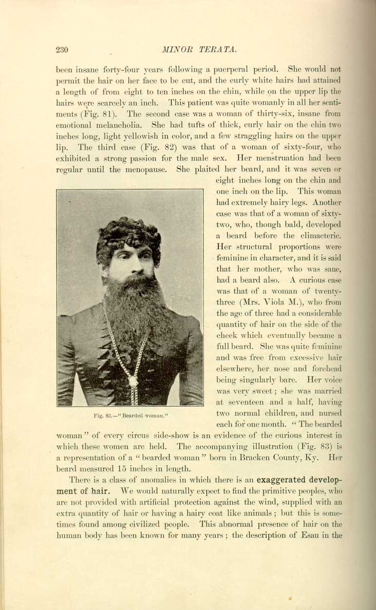
Fig. 83—"Bearded woman.''
[Description: Photograph of a woman with a full beard]
two normal children, and nursed each for one month. "The bearded woman'' of every circus side-show is an evidence of the curious interest in which these women are held. The accompanying illustration (Fig. 83) is a representation of a "bearded woman'' born in Bracken County, Ky. Her beard measured 15 inches in length.
There is a class of anomalies in which there is an exaggerated development of hair. We would naturally expect to find the primitive peoples, who are not provided with artificial protection against the wind, supplied with an extra quantity of hair or having a hairy coat like animals; but this is sometimes found among civilized people. This abnormal presence of hair on the human body has been known for many years; the description of Esau in the
In 1883 there was shown in England and France, afterward in America, a girl of seven named "Krao,'' a native of Indo-China. The whole body of this child was covered with black hair. Her face was of the prognathic type, and this, with her extraordinary prehensile powers of feet and lips, gave her the title of "Darwin's missing link.'' In 1875 there was exhibited in Paris, under the name of "l'homme-chien'' Adrien Jeftichew, a Russian
peasant of fifty-five, whose face, head, back, and limbs were covered with a brown hairy coat looking like wool and several centimeters long. The other parts of the body were also covered with hair, but less abundantly. This individual had a son of three, Theodore, who was hairy like himself.
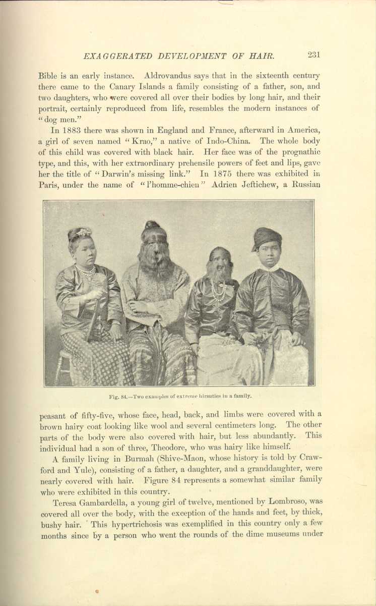
A family living in Burmah (Shive-Maon, whose history is told by Crawford and Yule), consisting of a father, a daughter, and a granddaughter, were nearly covered with hair. Figure 84 represents a somewhat similar family who were exhibited in this country.
Teresa Gambardella, a young girl of twelve, mentioned by Lombroso, was covered all over the body, with the exception of the hands and feet, by thick, bushy hair. This hypertrichosis was exemplified in this country only a few months since by a person who went the rounds of the dime museums under
Sometimes the hairy anomalies are but instances of nævus pilosus. The Indian ourang-outang woman examined at the office of the Lancet was an example of this kind. Hebra, Hildebrandt, Jablokoff, and Klein describe similar cases. Many of the older "wild men'' were individuals bearing extensive hairy moles.
Rayer remarks that he has seen a young man of sixteen who exhibited himself to the public under the name of a new species of wild man whose breast and back were covered with light brown hair of considerable length.
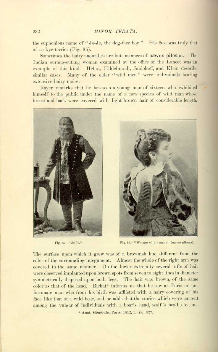
Fig. 85.—"Jo-Jo''
[Description: Photograph of a man whose face is covered in hair]

The surface upon which it grew was of a brownish hue, different from the color of the surrounding integument. Almost the whole of the right arm was covered in the same manner. On the lower extremity several tufts of hair were observed implanted upon brown spots from seven to eight lines in diameter symmetrically disposed upon both legs. The hair was brown, of the same color as that of the head. Bichat [6.52] informs us that he saw at Paris an unfortunate man who from his birth was afflicted with a hairy covering of his face like that of a wild boar, and he adds that the stories which were current among the vulgar of individuals with a boar's head, wolf's head, etc., undoubtedly
Duyse [6.54] reports a case of extensive hypertrichosis of the back in a girl aged nine years; her teeth were normal; there was pigmentation of the back and numerous pigmentary nevi on the face. Below each scapula there were tumors of the nature of fibroma molluscum. In addition to hairy nevi on the other parts of the body there was localized ichthyosis.
Ziemssen figures an interesting case of nævus pilosus resembling "bathing tights'' (Fig. 87). There were also present several benign tumors (fibroma molluscum) and numerous smaller nevi over the body. Schulz first observed the patient in 1878. This individual's name was Blake, and he stated that he was born with a large nævus spreading over the upper parts of the thighs and lower parts of the
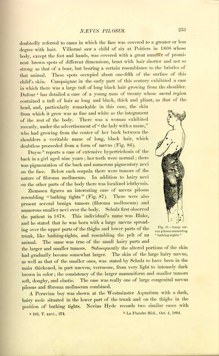
trunk, like bathing-tights, and resembling the pelt of an animal. The same was true of the small hairy parts and the larger and smaller tumors. Subsequently the altered portions of the skin had gradually become somewhat larger. The skin of the large hairy nævus, as well as that of the smaller ones, was stated by Schulz to have been in the main thickened, in part uneven, verrucose, from very light to intensely dark brown in color; the consistency of the larger mammiform and smaller tumors soft, doughy, and elastic. The case was really one of large congenital nævus pilosus and fibroma molluscum combined.
A Peruvian boy was shown at the Westminster Aquarium with a dark, hairy mole situated in the lower part of the trunk and on the thighs in the position of bathing tights. Nevins Hyde records two similar cases with
In some instances the hair and beard is of an enormous length. Erasmus Wilson of London saw a female of thirty-eight, whose hair measured 1.65 meters long. Leonard of Philadelphia speaks of a man in the interior of this country whose beard trailed on the ground when he stood upright, and measured 2.24 meters long. Not long ago there appeared the famous so-called "Seven Sutherland Sisters,'' whose hair touched the ground, and with whom nearly every one is familiar through a hair tonic which they extensively advertised. In Nature, January 9, 1892, is an account of a Percheron horse whose mane measured 13 feet and whose tail measured almost ten feet, probably
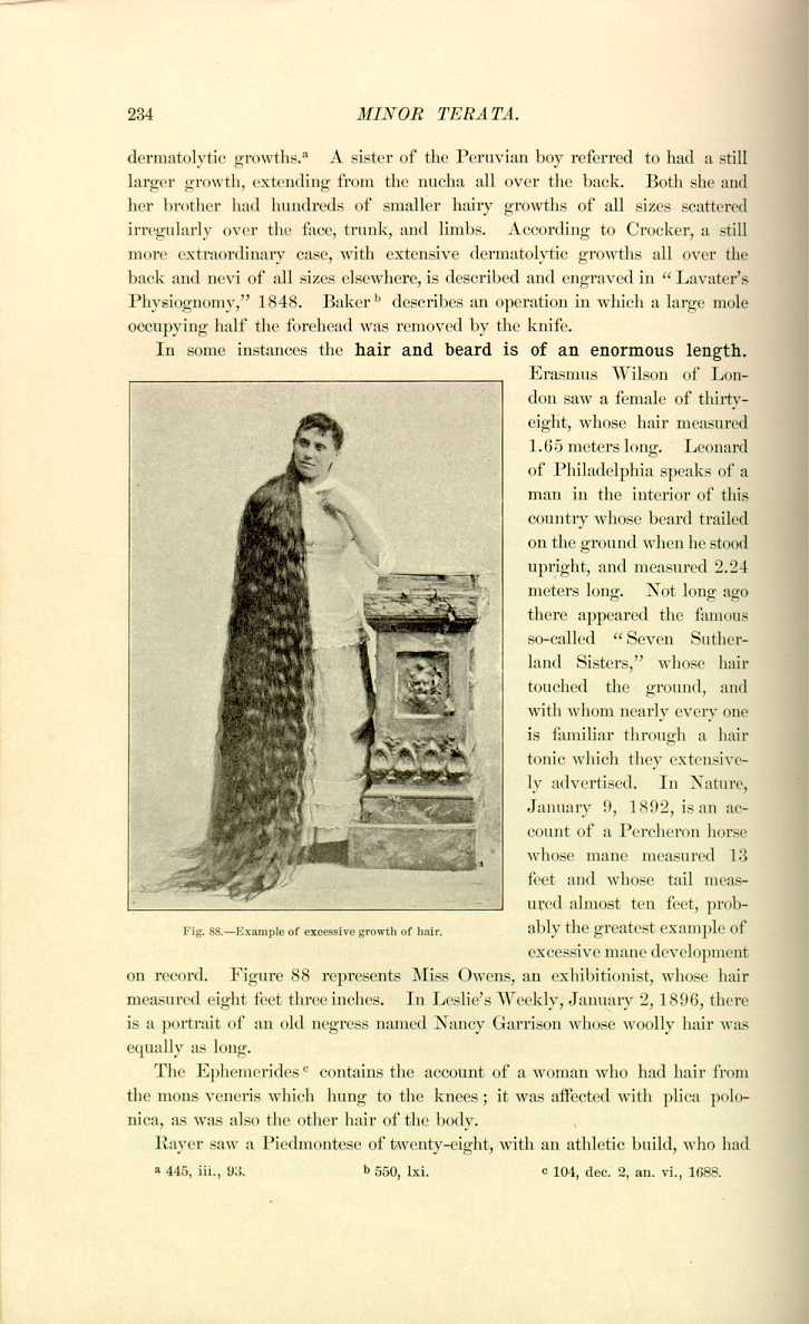
the greatest example of excessive mane development on record. Figure 88 represents Miss Owens, an exhibitionist, whose hair measured eight feet three inches. In Leslie's Weekly, January 2, 1896, there is a portrait of an old negress named Nancy Garrison whose woolly hair was equally as long.
The Ephemerides [6.57] contains the account of a woman who had hair from the mons veneris which hung to the knees; it was affected with plica polonica, as was also the other hair of the body.
Rayer saw a Piedmontese of twenty-eight, with an athletic build, who had
Certain pathologic conditions may give rise to accidental growths of hair. Boyer was accustomed to quote in his lectures the case of a man who, having an inflamed tumor in the thigh, perceived this part becoming covered in a short time with numerous long hairs. Rayer speaks of several instances of this kind. In one the part affected by a blister in a child of two became covered with hair. Another instance was that of a student of medicine, who after bathing in the sea for a length of time, and exposing himself to the hot sun, became affected with coppery patches, from which there sprang a growth of hair. Bricheteau, quoted by the same authority, speaks of a woman of twenty-four, having white skin and hair of deep black, who after a long illness occasioned by an affection analogous to marasmus became covered, especially on the back, breast, and abdomen, with a multitude of small elevations similar to those which appear on exposure to cold. These little elevations became brownish at the end of a few days, and short, fair, silky hair was observed on the summit of each, which grew so rapidly that the whole surface of the body with the exception of the hands and face became velvety. The hair thus evolved was afterward thrown out spontaneously and was not afterward reproduced.
Anomalies of the Color of the Hair.—New-born infants sometimes have tufts of hair on their heads which are perfectly white in color. Schenck speaks of a young man whose beard from its first appearance grew white. Young men from eighteen to twenty occasionally become gray; and according to Rayer, paroxysms of rage, unexpected and unwelcome news, diseases of the scalp such as favus, wounds of the head, habitual headache, over-indulgence of the sexual appetite, mercurial courses too frequently repeated, too great anxiety, etc., have been known to blanch the hair prematurely.
The well-accepted fact of the sudden changing of the color of the hair from violent emotions or other causes has always excited great interest, and many ingenious explanations have been devised to account for it. There is a record in the time of Charles V. of a young man who was committed to prison in 1546 for seducing his girl companion, and while there was in great fear and grief, expecting a death-sentence from the Emperor the next day. When brought before his judge, his face was wan and pale and his hair and beard gray, the change having taken place in the night. His beard was filthy with drivel, and the Emperor, moved by his pitiful condition, pardoned him. There was a clergyman [6.58] of Nottingham whose daughter at the age of thirteen experienced a change from jet-blackness of the hair to white in a single night, but this was confined to a spot on the back of the head 1 1/2
Voigtel mentions the occurrence of canities almost suddenly. Bichat had a personal acquaintance whose hair became almost entirely gray in consequence of some distressing news that reached him. Cassan [6.59] records a similar case. According to Rayer, a woman by the name of Perat, summoned before the Chamber of Peers to give evidence in the trial of the assassin Louvel, was so much affected that her hair became entirely white in a single night Byron makes mention of this peculiar anomaly in the opening stanzas of the "Prisoner of Chillon:''—
Nor grew it white
In a single night.
As men's have grown from sudden fears.''
The commentators say that Byron had reference to Ludovico Sforza and others. The fact of the change is asserted of Marie Antoinette, the wife of Louis XVI., though in not quite so short a period, grief and not fear being the cause. Ziemssen cites Landois' case of a compositor of thirty-four who was admitted to a hospital July 9th with symptoms of delirium tremens; until improvement began to set in (July 13th) he was continually tormented by terrifying pictures of the imagination. In the night preceding the day last mentioned the hair of the head and beard of the patient, formerly blond, became gray. Accurate examination by Landois showed the pigment contents of the hair to be unchanged, and led him to believe that the white color was solely due to the excessive development of air-bubbles in the hair shaft. Popular belief brings the premature and especially the sudden whitening into connection with depressing mental emotions. We might quote the German expression—"Sich graue Haare etwas wachsen lassen'' ("To worry one's self gray''). Brown-Séquard observed on several occasions in his own dark beard hairs which had turned white in a night and which he epilated. He closes his brief communication on the subject with the belief that it is quite possible for black hair to turn white in one night or even in a less time, although Hebra and Kaposi discredit sudden canities (Duhring). Raymond and Vulpian [6.60] observed a lady of neurotic type whose hair during a severe paroxysm of neuralgia following a mental strain changed color in five hours over the entire scalp except on the back and sides; most of the hair changed from black to red, but some to quite white, and in two days
Dewees [6.61] reports a case of puerperal convulsions in a patient under his care which was attended with sudden canities. From 10 A. M. to 4 P. M. 50 ounces of blood were taken. Between the time of Dr. Dewees' visits, not more than an hour, the hair anterior to the coronal suture turned white. The next day it was less light, and in four or five days was nearly its natural color. He also mentions two cases of sudden blanching from fright.
Fowler [6.62] mentions the case of a healthy girl of sixteen who found one morning while combing her hair, which was black, that a strip the whole length of the back hair was white, starting from a surface about two inches square around the occipital protuberance. Two weeks later she had patches of ephelis over the whole body.
Prentiss, in Science, October 3, 1890, has collected numerous instances of sudden canities, several of which will be given:—
"In the Canada Journal of Medical Science, 1882, p. 113, is reported a case of sudden canities due to business-worry. The microscope showed a great many air-vesicles both in the medullary substance and between the medullary and cortical substance.
"In the Boston Medical and Surgical Journal, 1851, is reported a case of a man thirty years old, whose hair `was scared' white in a day by a grizzly bear. He was sick in a mining camp, was left alone, and fell asleep. On waking he found a grizzly bear standing over him.
"A second case is that of a man of twenty-three years who was gambling in California. He placed his entire savings of $1100 on the turn of a card. He was under tremendous nervous excitement while the cards were being dealt. The next day his hair was perfectly white.
"In the same article is the statement that the jet-black hair of the Pacific Islanders does not turn gray gradually, but when it does turn it is sudden, usually the result of fright or sudden emotions.''
D'Alben, quoted by Fournier, [6.63] describes a young man of twenty-four, an officer in the regiment of Touraine in 1781, who spent the night in carnal dissipation with a mulatto, after which he had violent spasms, rendering flexion of the body impossible. His beard and hair on the right side of the body was found as white as snow, the left side being unchanged. He appeared before the Faculté de Montpelier, and though cured of his nervous symptoms his hair was still white, and no suggestion of relief was offered him.
Louis of Bavaria, who died in 1294, on learning of the innocence of his
De Schweinitz [6.64] speaks of a well-formed and healthy brunette of eighteen in whom the middle portion of the cilia of the right upper eyelid and a number of the hairs of the lower lid turned white in a week. Both eyes were myopic, but no other cause could be assigned. Another similar case is cited by Hirshberg, [6.65] and the authors have seen similar cases. Thornton of Margate records the case of a lady in whom the hair of the left eyebrow and eyelashes began to turn white after a fortnight of sudden grief, and within a week all the hair of these regions was quite white and remained so. No other part was affected nor was there any other symptom. After a traumatic ophthalmitis of the left and sympathetic inflammation of the right eye in a boy of nine, Schenck observed that a group of cilia of the right upper lid and nearly all the lashes of the upper lid of the left eye, which had been enucleated, turned silvery-white in a short time. Ludwig has known the eyelashes to become white after small-pox. Communications are also on record of local decolorization of the eyebrows and lashes in neuralgias of isolated branches of the trigeminus, especially of the supraorbital nerve.
Temporary and Partial Canities.—Of special interest are those cases in which whiteness of the hair is only temporary. Thus, Compagne mentions a case in which the black hair of a woman of thirty-six began to fade on the twenty-third day of a malignant fever, and on the sixth day following was perfectly white, but on the seventh day the hairs became darker again, and on the fourteenth day after the change they had become as black as they were originally. Wilson records a case in which the hair lost its color in winter and regained it in summer. Sir John Forbes, according to Crocker, had gray hair for a long time, then suddenly it all turned white, and after remaining so for a year it returned to its original gray.
Grayness of the hair is sometimes only partial. According to Crocker an adult whose hair was generally brown had a tuft of white hair over the temple, and several like cases are on record. Lorry tells us that grayness of one side only is sometimes occasioned by severe headache. Hagedorn has known the beard to be black in one place and white in another. Brandis mentions the hair becoming white on one side of the face while it continued of its former color on the other. Rayer quotes cases of canities of the whole of one side of the body.
Richelot observed white mottling of hair in a girl sick with chlorosis.
Michelson of Königsberg describes a curious case in a barrister of twenty-three affected with partial canities. In the family of both parents there was stated to be congenital premature canities, and some white hairs had been observed even in childhood. In the fifteenth year, after a grave attack of scarlet fever, the hair to a great extent fell out. The succeeding growth of hair was stated to have been throughout lighter in tissue and color and fissured at the points. Soon after bunches of white hair appeared on the occiput, and in the succeeding years small patches of decolored hairs were observed also on the anterior and lateral portions of the scalp. In the spring of 1880 the patient exhibited signs of infiltration of the apex of the right lung, and afterward a violent headache came on. At the time of the report the patient presented the appearance shown in Figure 89. The complexion was delicate throughout, the eyelashes and eyelids dark brown, the moustache and whiskers blond, and in the latter were a few groups of white hair. The white patches were chiefly on the left side of the head. The hairs growing on
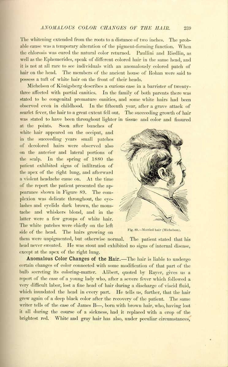
Fig.89.—Mottled hair (Michelson).
[Description: Drawing of man's head with mottled hair, rear view]
them were unpigmented, but otherwise normal. The patient stated that his head never sweated. He was stout and exhibited no signs of internal disease, except at the apex of the right lung.
Anomalous Color Changes of the Hair.—The hair is liable to undergo certain changes of color connected with some modification of that part of the bulb secreting its coloring-matter. Alibert, quoted by Rayer, gives us a report of the case of a young lady who, after a severe fever which followed a very difficult labor, lost a fine head of hair during a discharge of viscid fluid, which inundated the head in every part. He tells us, further, that the hair grew again of a deep black color after the recovery of the patient. The same writer tells of the case of James B—, born with brown hair, who, having lost it all during the course of a sickness, had it replaced with a crop of the brightest red. White and gray hair has also, under peculiar circumstances,
A very singular case, published early in the century, was that of a woman whose hair, naturally fair, assumed a tawny red color as often as she was affected with a certain fever, and returned to its natural hue as soon as the symptoms abated. [6.67] Villermé *[302] alludes to the case of a young lady, sixteen years of age, who had never suffered except from trifling headaches, and who, in the winter of 1817, perceived that the hair began to fall out from several parts of her head, so that before six months were over she became entirely bald. In the beginning of January, 1819, her head became covered with a kind of black wool over those places that were first denuded, and light brown hair began to develop from the rest of the scalp. Some of this fell out again when it had grown from three to four inches; the rest changed color at different distances from its end and grew of a chestnut color from the roots. The hair, half black, half chestnut, had a very singular appearance.
Alibert and Beigel relate cases of women with blond hair which all came off after a severe fever (typhus in one case), and when it grew again it was quite black. Alibert also saw a young man who lost his brown hair after an illness, and after restoration it became red. According to Crocker, in an idiotic girl of epileptic type (in an asylum at Edinburgh), with alternating phases of stupidity and excitement, the hair in the stupid phase was blond and in the excited condition red. The change of color took place in the course of two or three days, beginning first at the free ends, and remaining of the same tint for seven or eight days. The pale hairs had more air-spaces than the darker ones. There was much structural change in the brain and spinal cord. Smyly of Dublin reported a case of suppurative disease of the temporal bone, in which the hair changed from a mouse-color to a reddish-brown; and Squire records a congenital case in a deaf mute, in whom the hair on the left side was in light patches of true auburn and dark patches of dark brown like a tortoise-shell cap; on the other side the hair was a dark brown. Crocker mentions the changes which have occurred in rare instances after death from dark brown to red.
Chemic colorations of various tints occur. Blue hair is seen in workers in cobalt mines and indigo works; green hair in copper smelters; deep red-brown hair in handlers of crude anilin; and the hair is dyed a purplish-brown whenever chrysarobin applications used on a scalp come in contact with an alkali, as when washed with soap. Among such cases in older
Many curious causes are given for alopecia. Gilibert and Merlet [6.68] mention sexual excess; Marcellus Donatus [6.69] gives fear; the Ephemerides speaks of baldness from fright; and Leo Africanus, in his description of Barbary, describes endemic baldness. Neyronis [6.70] makes the following observation: A man of seventy-three, convalescent from a fever, one morning, about six months after recovery perceived that he had lost all his hair, even his eyelashes, eyebrows, nostril-hairs, etc. Although his health continued good, the hair was never renewed.
The principal anomalies of the nails observed are absence, hypertrophy, and displacement of these organs. Some persons are born with finger-nails and toe-nails either very rudimentary or entirely absent; in others they are of great length and thickness. The Chinese nobility allow their finger-nails to grow to a great length and spend much time in the care of these nails. Some savage tribes have long and thick nails resembling the claws of beasts, and use them in the same way as the lower animals. There is a description of a person with finger-nails that resembled the horns of a goat. [6.71]
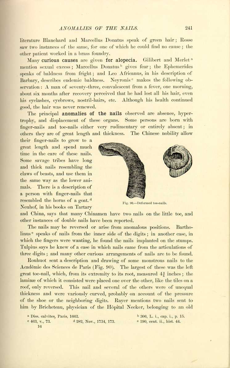
Fig. 90.—Deformed toe-nails.
[Description: Drawing of deformed toe-nails]
Neuhof, in his books on Tartary and China, says that many Chinamen have two nails on the little toe, and other instances of double nails have been reported.
The nails may be reversed or arise from anomalous positions. Bartholinus [6.72] speaks of nails from the inner side of the digits; in another case, in which the fingers were wanting, he found the nails implanted on the stumps. Tulpius says he knew of a case in which nails came from the articulations of three digits; and many other curious arrangements of nails are to be found.
Rouhuot sent a description and drawing of some monstrous nails to the Académie des Sciences de Paris (Fig. 90). The largest of these was the left great toe-nail, which, from its extremity to its root, measured 4 3/4 inches; the laminæ of which it consisted were placed one over the other, like the tiles on a roof, only reversed. This nail and several of the others were of unequal thickness and were variously curved, probably on account of the pressure of the shoe or the neighboring digits. Rayer mentions two nails sent to him by Bricheteau, physician of the Hôpital Necker, belonging to an old
Musaeus [6.73] gives an account of the nails of a girl of twenty, which grew to such a size that some of those of the fingers were five inches in length. They were composed of several layers, whitish interiorly, reddish-gray on the exterior, and full of black points. These nails fell off at the end of four months and were succeeded by others. There were also horny laminæ on the knees and shoulders and elbows which bore a resemblance to nails, or rather talons. They were sensitive only at the point of insertion into the skin. Various other parts of the body, particularly the backs of the hands, presented these horny productions. One of them was four inches in length. This horny growth appeared after small-pox. Ash, in the Philosophical Transactions, records a somewhat similar case in a girl of twelve.
Anomalies of the Teeth.—Pliny, Colombus, van Swieten, Haller, Marcellus Donatus, Baudelocque, Soemmering, and Gardien all cite instances in which children have come into the world with several teeth already erupted. Haller *[400] has collected 19 cases of children born with teeth. Polydorus Virgilus describes an infant who was born with six teeth. Some celebrated men are supposed to have been born with teeth; Louis XIV. was accredited with having two teeth at birth. Bigot, a physician and philosopher of the sixteenth century; Boyd, the poet; Valerian, Richard III., as well as some of the ancient Greeks and Romans, were reputed to have had this anomaly. The significance of the natal eruption of teeth is not always that of vigor, as many of the subjects succumb early in life. There were two cases typical of fetal dentition shown before the Académie de Médecine de Paris. One of the subjects had two middle incisors in the lower jaw and the other had one tooth well through. Levison [6.74] saw a female born with two central incisors in the lower jaw.
Thomas [6.75] mentions a case of antenatal development of nine teeth. Puech, Mattei, Dumas, Belluzi, and others report the eruption of teeth in the newborn. In Dumas' case the teeth had to be extracted on account of ulceration of the tongue.
Edentulousness.—We have already noticed the association of congenital alopecia with edentulousness, but, strange to say, Magitot has remarked that "l'homme-chien,'' was the subject of defective dentition. Borellus found atrophy of all the dental follicles in a woman of sixty who never had possessed any teeth. Fanton-Touvet saw a boy of nine who had never had teeth, and Fox a woman who had but four in both jaws; Tomes cites several similar instances. Hutchinson [6.86] speaks of a child who was perfectly edentulous as to temporary teeth, but who had the permanent teeth duly and fully erupted. Guilford [6.87] describes a man of forty-eight, who was edentulous from birth, who also totally lacked the sense of smell, and was almost without the sense
The anomalies of excessive dentition are of several varieties, those of simple supernumerary teeth, double or triple rows, and those in anomalous positions. Ibbetson saw a child with five incisors in the inferior maxillary bone, and Fanton-Touvet describes a young lady who possessed five large incisors of the first dentition in the superior maxilla. Rayer [6.88] notes a case of dentition of four canines, which first made their appearance after pain for eight days in the jaws and associated with convulsions. In an Ethiopian Soemmering has seen one molar too many on each side and in each jaw. Ploucquet and Tesmer have seen five incisors and Fanchard six. Many persons have the supernumerary teeth parallel with their neighbors, anteriorly or posteriorly. Costa [6.89] reports a case in which there were five canine teeth in the upper jaw, two placed laterally on either side, and one on the right side behind the other two. The patient was twenty-six years of age, well formed and in good health.
In some cases there is fusion of the teeth. Pliny, Bartholinus, and Melanthon pretend to have seen the union of all the teeth, making a continuous mass. In the "Musée de l'école dentaire de Paris'' there are several milk-teeth, both of the superior and inferior maxilla, which are fused together. Bloch cites a case in which there were two rows of teeth in the superior maxilla. Hellwig *[414] has observed three rows of teeth, and the Ephemerides contain an account of a similar anomaly.
Extraoral Dentition.—Probably the most curious anomaly of teeth is that in which they are found in other than normal positions. Albinus speaks of teeth in the nose and orbit; Borellus, in the palate; Fabricius Hildanus, *[334] under the tongue; Schenck, from the palate; and there are many similar modern records. Heister in 1743 wrote a dissertation on extraoral teeth. The following is a recent quotation: [6.90]—
"In the Norsk Magazin fur Laegevidenskaben, January, 1895, it is reported that Dr. Dave, at a meeting of the Medical Society in Christiania, showed a tooth removed from the nose of a woman aged fifty-three. The patient had consulted him for ear-trouble, and the tooth was found accidentally during the routine examination. It was easily removed, having
Delpech saw a young man in 1829 who had an opening in the palatine vault occasioned by the extraction of a tooth. This opening communicated with the nasal fossa by a fracture of the palatine and maxillary bones; the employment of an obturator was necessary. It is not rare to see teeth, generally canine, make their eruption from the vault of the palate; and these teeth are not generally supernumerary, but examples of vice and deviation of position. Fanton-Touvet, however, gives an example of a supernumerary tooth implanted in the palatine arch. Branch [6.91] describes a little negro boy who had two large teeth in the nose; his dentition was otherwise normal, but a portion of the nose was destroyed by ulceration. Roy [6.92] describes a Hindoo lad of fourteen who had a tooth in the nose, supposed to have been a tumor. It was of the canine type, and was covered with enamel to the junction with the root, which was deeply imbedded in the side and upper part of the antrum. The boy had a perfect set of permanent teeth and no deformity, swelling, or cystic formation of the jaw. This was clearly a case of extrafollicular development and eruption of the tooth in an anomalous position, the peculiarity being that while in other similar cases the crown of the tooth shows itself at the floor of the nasal cavity from below upward, in this instance the dental follicle was transposed, the eruption being from above downward. Hall [6.93] cites an instance in which the right upper canine of a girl erupted in the nose. The subject showed marked evidence of hereditary syphilis. Carver [6.94] describes a child who had a tooth growing from the lower right eyelid. The number of deciduous teeth was perfect; although this tooth was canine it had a somewhat bulbulous fang.
Of anomalies of the head the first to be considered will be the anencephalous monsters who, strange to say, have been known to survive birth. Clericus [6.95] cites an example of life for five days in a child without a cerebrum.
Fraser [6.102] mentions a brother and sister, aged twenty and thirty, respectively, who from birth had exhibited signs of defective development of the cerebellum. They lacked power of coordination and walked with a drunken, staggering gait; they could not touch the nose with the finger when their eyes were shut, etc. The parents of these unfortunate persons were perfectly healthy, as were the rest of their family. Cruveilhier cites a case of a girl of eleven who had absolutely no cerebellum, with the same symptoms which are characteristic in such cases. There is also recorded the history of a man [6.103] who was deficient in the corpus callosum; at the age of sixty-two, though of feeble intelligence, he presented no signs of nervous disorder. Claude Bernard made an autopsy on a woman who had no trace of olfactory lobes, and after a minute inquiry into her life he found that her sense of smell had been good despite her deficiency.
Buhring relates the history of a case somewhat analogous to viability of anencephalous monsters. It was a bicephalous child that lived thirty-two hours after he had ligated one of its heads. [6.104]
Ward [6.105] mentions an instance of congenital absence of the corpora callosum.
Diminution in volume of the head is called microcephaly. Probably the most remarkable case on record is that mentioned by Lombroso. The
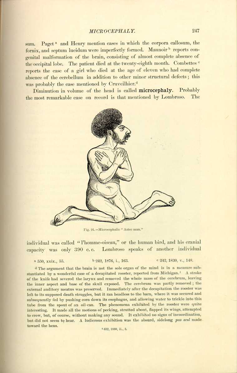
Fig. 91.—Microcephalic "Aztec man.''
[Description: Drawing of "Aztec man"]
individual was called "l'homme-oiseau,'' or the human bird, and his cranial capacity was only 390 c. c. Lombroso speaks of another individual
Two creatures of celebrity were Maximo and Bartola, who for twenty-five years have been shown in America and in Europe under the name of the "Aztecs'' or the "Aztec children'' (Fig. 91). They were male and female and very short, with heads resembling closely the bas-reliefs on the ancient Aztec temples of Mexico. Their facial angle was about 45°, and they had jutting lips and little or no chin. They wore their hair in an enormous bunch to magnify the deformity. These curiosities were born in Central America and were possibly half Indian and Negro. They were little better than idiots in point of intelligence.
Figure 92 represents a microcephalic youth known as the "Mexican wild boy,'' who was shown with the Wallace circus.
Virchow [6.110] exhibited a girl of fourteen whose face was no larger than that of a new-born child, and whose head was scarcely as large as a man's fist. Magitot reported a case of a microcephalic woman of thirty who weighed 70 pounds.
Hippocrates and Strabonius both speak of head-binding as a custom inducing artificial microcephaly, and some tribes of North American Indians still retain this custom.
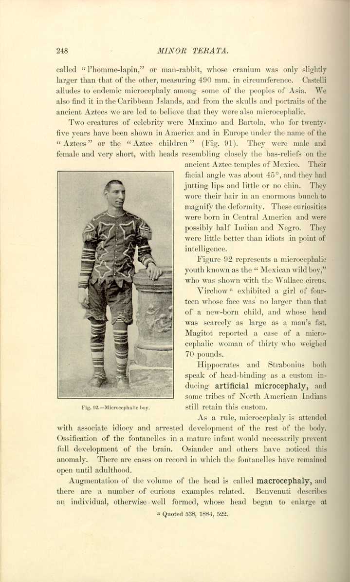
Fig.92.—Microcephalic boy.
[Description: Photograph of microcephalic boy]
As a rule, microcephaly is attended with associate idiocy and arrested development of the rest of the body. Ossification of the fontanelles in a mature infant would necessarily prevent full development of the brain. Osiander and others have noticed this anomaly. There are cases on record in which the fontanelles have remained open until adulthood.
Augmentation of the volume of the head is called macrocephaly, and there are a number of curious examples related. Benvenuti describes an individual, otherwise well formed, whose head began to enlarge at
Fournier [6.112] speaks of a cranium in the cabinet of the Natural History Museum of Marseilles of a man by the name of Borghini, who died in 1616. At the time he was described he was fifty years old, four feet in height; his head measured three feet in circumference and one foot in height. There was a proverb in Marseilles, "Apas maï de sen que Borghini,'' meaning in the local dialect, "Thou hast no more wit than Borghini.'' This man, whose fame became known all over France, was not able, as he grew older, to maintain the weight of his head, but carried a cushion on each shoulder to prop it up. Fournier also quotes the history of a man who died in the same city in 1807 at the age of sixty-seven. His head was enormous, and he never lay on a bed for thirty years, passing his nights in a chair, generally reading or writing. He only ate once in twenty-four or thirty hours, never warmed himself, and never used warm water. His knowledge was said to have been great and encyclopedic, and he pretended never to have heard the proverb of Borghini. There is related the account of a Moor, who was seen in Tunis early in this century, thirty-one years of age, of middle height, with a head so prodigious in dimensions that crowds flocked after him in the streets. His nose was quite long, and his mouth so large that he could eat a melon as others would an apple. He was an imbecile. William Thomas Andrews was a dwarf seventeen years old, whose head measured in circumference 35 inches; from one external auditory meatus to another, 27 1/4 inches; from the chin over the cranial summit to the suboccipital protuberance, 37 1/2 inches; the distance from the chin to the pubes was 20 inches; and from the pubes to the soles of the feet, 16; he was a monorchid. [6.113] James Cardinal, who died in Guy's Hospital in 1825, and who was so celebrated for the size of his head, only measured 32 1/2 inches in head-circumference.
The largest healthy brains on record, that is, of men of prominence, are those of Cuvier, weighing 64 1/3 ounces; [6.114] of Daniel Webster, weighing 63 3/4 ounces (the circumference of whose head was 23 3/4 inches); [6.115] of Abercrombie, weighing 63 ounces, and of Spurzheim, weighing 55 1/16 ounces. Byron and Cromwell had abnormally heavy brains, showing marked evidence of disease.
A curious instance in this connection is that quoted by Pigné, [6.116] who gives an account of a double brain found in an infant. Keen reports finding a fornix which, instead of being solid from side to side, consisted of two lateral halves with a triangular space between them.
When the augmentation of the volume of the cranium is caused by an abundant quantity of serous fluid the anomaly is known as hydrocephaly.
There is a record [6.117] of a curious monster born of healthy half-caste African parents. The deformity was caused by

Fig 93.—Hydrocephalic child.
[Description: Photograph of hydrocephalic child]
a deficiency of osseous material of the bones of the head. There was considerable arrest of development
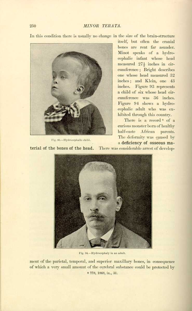
Fig. 94. Hydrocephaly in an adult.
[Description: Photograph of hydrocephalic adult]
of the parietal, temporal, and superior maxillary bones, in consequence of which a very small amount of the cerebral substance could be protected by
Occasionally a deficiency in the osseous material of the cranium or an abnormal dilatation of the fontanelles

gives rise to a hernia of the meninges, which, if accompanied by cerebrospinal fluid in any quantity, causes a large and peculiarly shaped tumor called meningocele (Fig. 96). If there is a protrusion of brain-substance itself, a condition known as hernia cerebri results.
Complete absence of the inferior maxilla is much rarer in man than in animals. Nicolas and Prenant have described a curious case of this anomaly in a sheep. Gurlt has named subjects presenting the total or partial absence of the inferior maxilla, agnathes or hemiagnathes. Simple atrophy of the inferior maxilla has been seen in man as well as in the lower animals, but is much less frequent than atrophy of the superior maxilla. Langenbeck reports the case of a young man who had the inferior maxilla so atrophied
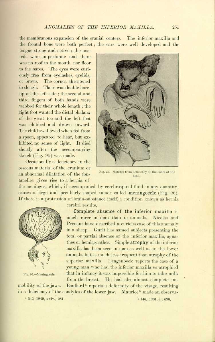
Fig. 96.—Meningocele.
[Description: Drawing of tumor of the head]
that in infancy it was impossible for him to take milk from the breast. He had also almost complete immobility of the jaws. Boullard [6.118] reports a deformity of the visage, resulting in a deficiency of the condyles of the lower jaw. Maurice [6.119] made an observation
Exaggerated prominence of the maxillaries is called prognathism; that of the superior maxilla is seen in the North American Indians. Inferior prognathism is observed in man as well as in animals. The bull-dog, for example, displays this, but in this instance the deformity is really superior brachygnathism, the superior maxilla being arrested in development.
Congenital absence of the nose is a very rare anomaly. Maisonneuve has seen an example in an individual in which, in place of the nasal appendix, there was a plane surface perforated by two small openings a little less than one mm. in diameter and three mm. apart.
Exaggeration in volume of the nose is quite frequent. Ballonius *[185] speaks of a nose six times larger than ordinary. Viewing the Roman celebrities, we find that Numa, to whom was given the surname Pompilius, had a nose which measured six inches. Plutarch, Lyourgus, and Solon had a similar enlargement, as had all the kings of Italy except Tarquin the Superb.
Early in the last century a man, Thomas Wedders (or Wadhouse), with a nose 7 1/2 inches long, was exhibited throughout Yorkshire. This man expired
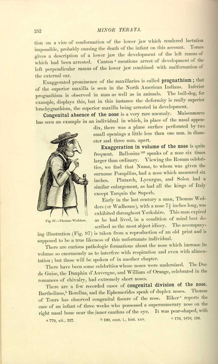
Fig. 97.—Thomas Wedders.
[Description: Drawing of Thomas Wedders]
as he had lived, in a condition of mind best described as the most abject idiocy. The accompanying illustration (Fig. 97) is taken from a reproduction of an old print and is supposed to be a true likeness of this unfortunate individual.
There are curious pathologic formations about the nose which increase its volume so enormously as to interfere with respiration and even with alimentation; but these will be spoken of in another chapter.
There have been some celebrities whose noses were undersized. The Duc de Guise, the Dauphin d'Auvergne, and William of Orange, celebrated in the romances of chivalry, had extremely short noses.
There are a few recorded cases of congenital division of the nose. Bartholinus, [6.121] Borellus, and the Ephemerides speak of duplex noses. Thomas of Tours has observed congenital fissure of the nose. Riker [6.122] reports the case of an infant of three weeks who possessed a supernumerary nose on the right nasal bone near the inner canthus of the eye. It was pear-shaped, with
Anomalies in size of the mouth are not uncommon. Fournier quotes the history of a man who had a mouth so large that when he opened it all his back teeth could be seen. There is a history of a boy of seventeen [6.126] who had a preternaturally-sized mouth, the transverse diameter being 6 1/2 inches. The mother claimed that the boy was born with his foot in his mouth and to this fact attributed his deformity. The negro races are noted for their large mouths and thick lips. A negro called "Black Diamond,'' recently exhibited in Philadelphia, could put both his fists in his mouth.
Morgan [6.127] reports two cases of congenital macrostomaaccompanied by
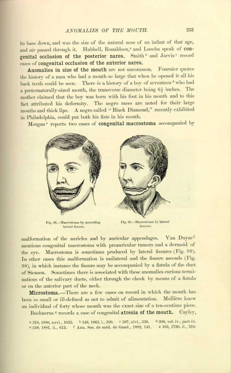
Fig. 98.—Macrostoma by ascending
lateral fissure.
[Description: Drawing of macrrostoma]

Fig. 99.—Macrostoma by lateral
fissures.
[Description: Drawing of macrostoma by lateral fissures]
malformation of the auricles and by auricular appendages. Van Duyse [6.128] mentions congenital macrostoma with preauricular tumors and a dermoid of the eye. Macrostoma is sometimes produced by lateral fissures (Fig. 99). In other cases this malformation is unilateral and the fissure ascends (Fig. 98), in which instance the fissure may be accompanied by a fistula of the duct of Stensen. Sometimes there is associated with these anomalies curious terminations of the salivary ducts, either through the cheek by means of a fistula or on the anterior part of the neck.
Microstoma.—There are a few cases on record in which the mouth has been so small or ill-defined as not to admit of alimentation. Mollière knew an individual of forty whose mouth was the exact size of a ten-centime piece.
Buchnerus [6.129] records a case of congenital atresia of the mouth. Cayley,
Anomalies of the Lips.—The aboriginal tribes are particularly noted for their large and thick lips, some of which people consider enormous lips signs of adornment. Elephantiasis or other pathologic hypertrophy of the labial tissues can produce revolting deformity, such as is seen in Figure 100, representing an individual who was exhibited several years ago in Philadelphia. We have in English the expression, "pulling a long lip.'' Its origin is said to date back to a semimythical hero of King Arthur's time, who, "when sad at heart and melancholic,'' would let one of his lips drop below his waist, while he turned the other up like a cap on his head.
Blot [6.132] records a case of monstrous congenital hypertrophy of the superior lip in an infant of eight months. Buck [6.133] successfully treated by surgical operations a case of congenital hypertrophy of the under lip, and Detmold [6.134] mentions a similar result in a young lady with hypertrophy of the lip and lower part of the nose. Murray [6.135] reports an undescribed malformation of the lower lip occurring in one family.
Hare-lip may be unilateral or double, and may or may not include the palatine
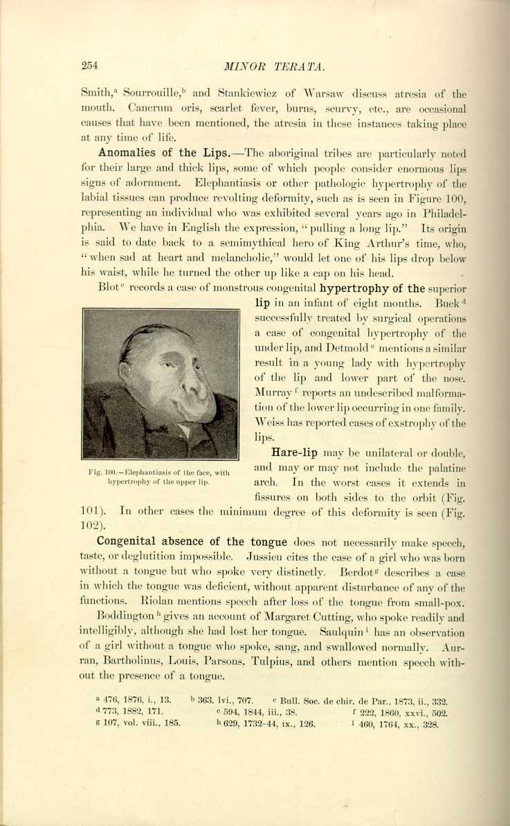
arch. In the worst cases it extends in fissures on both sides to the orbit (Fig. 101). In other cases the minimum degree of this deformity is seen (Fig. 102).
Congenital absence of the tongue does not necessarily make speech, taste, or deglutition impossible. Jussieu cites the case of a girl who was born without a tongue but who spoke very distinctly. Berdot [6.136] describes a case in which the tongue was deficient, without apparent disturbance of any of the functions. Riolan mentions speech after loss of the tongue from small-pox.
Boddington [6.137] gives an account of Margaret Cutting, who spoke readily and intelligibly, although she had lost her tongue. Saulquin [6.138] has an observation of a girl without a tongue who spoke, sang, and swallowed normally. Aurran, Bartholinus, Louis, Parsons, Tulpius, and others mention speech without the presence of a tongue.
Jurist [6.140] records such abnormal mobility of the tongue that the patient was able to project the tongue into the nasopharynx. Wherry and Winslow record similar instances.
There have been individuals with bifid tongues, after the normal type of serpents and saurians, and others who possessed a supernumerary tongue. Rev. Henry Wharton, Chaplain to Archbishop Sancroft, in his journal, written in the seventeenth century, says that he was born with two tongues and
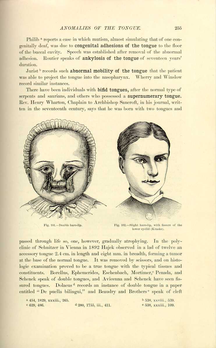
Fig. 101.—Double hare-lip.
[Description: Drawing of a child with a double hare-lip]

passed through life so, one, however, gradually atrophying. In the polyclinic of Schnitzer in Vienna in 1892 Hajek observed in a lad of twelve an accessory tongue 2.4 cm. in length and eight mm. in breadth, forming a tumor at the base of the normal tongue. It was removed by scissors, and on histologic examination proved to be a true tongue with the typical tissues and constituents. Borellus, Ephemerides, Eschenbach, Mortimer, [6.141] Penada, and Schenck speak of double tongues, and Avicenna and Schenck have seen fissured tongues. Dolaeus [6.142] records an instance of double tongue in a paper entitled "De puella bilingui,'' and Beaudry and Brothers [6.143] speak of cleft
In some cases there is marked augmentation of the volume of the tongue. Fournier [6.145] has seen a juggler with a tongue so long that he could extrude it six inches from his mouth. He also refers to a woman in Berlin with a long tongue, but it was thinner than that of a cat. When she laughed it hung over her teeth like a curtain, and was always extremely cold to the touch. In the same article there is a description of a man with a very long neck who could touch his tongue to his chest without reclining his head. Congenital and acquired hypertrophy of the tongue will be discussed later.
Amatus Lusitanus [6.146] and Portal [6.147] refer to the presence of hair on the tongue, and later there was an account of a medical student [6.148] who complained of dyspepsia and a sticky sensation in the mouth. On examination a considerable growth of hair was found on the surface of the tongue. The hairs would be detached in vomiting but would grow again, and when he was last seen they were one inch long. Such are possibly nevoid in formation.
The ordinary anomalies of the palate are the fissures, unilateral, bilateral, median, etc.: they are generally associated with hare-lip. The median fissure commencing between the middle incisors is quite rare.
Many curious forms of obturator or artificial palate are employed to remedy congenital defects. Sercombe [6.149] mentions a case in which destruction of the entire palate was successfully relieved by mechanical means. In some instances among the lower classes these obturators are simple pieces of wood, so fashioned as to fit into the palatine cleft, and not infrequently the obturator has been swallowed, causing obstruction of the air-passages or occluding the esophagus.
Abnormalism of the Uvula.—Examples of double uvula are found in the older writers, and Hagendorn speaks of a man who was born without a uvula. The Ephemerides and Salmuth describe uvulæ so defective as to be hardly noticeable. Bolster, Delius, Hodges, Mackenzie of Baltimore, Orr, Riedel, Schufeldt, and Tidyman are among observers reporting bifurcated and double uvula, and they are quite common. Ogle [6.150] records instances of congenital absence of the uvula.
Anomalies of the Epiglottis.—Morgagni mentions a man without an epiglottis who ate and spoke without difficulty. He thought the arytenoids were so strongly developed that they replaced the functions of the missing organ. Enos of Brooklyn in 1854 reported absence of the epiglottis without interference with deglutition. [6.151] Manifold [6.152] speaks of a case of bifurcated epiglottis. Debloisi [6.153] records an instance of congenital web of the
Instances of duplication of the epiglottis attended witha species of double voice possess great interest. French [6.155] described a man of thirty, by occupation a singer and contortionist, who became possessed of an extra voice when he was sixteen. In high and falsetto tones he could run the scale from A to F in an upper and lower range. The compass of the low voice was so small that he could not reach the high notes of any song with it, and in singing he only used it to break in on the falsetto and produce a sensation. He was supposed to possess a double epiglottis. [6.156]
Roe [6.157] describes a young lady who could whistle at will with the lower part of her throat and without the aid of her lips. Laryngeal examination showed that the fundamental tones were produced by vibrations of the edges of the vocal cords, and the modifications were effected by a minute adjustment of the ventricular bands, which regulated the laryngeal opening above the cord, and pressing firmly down closed the ventricle and acted as a damper preventing the vibrations of the cords except in their middle third. Morgan in the same journal mentions the case of a boy of nineteen, who seemed to be affected with laryngeal catarrh, and who exhibited distinct diphthongia. He was seen to have two glottic orifices with associate bands. The treatment was directed to the catarrh and consequent paresis of the posterior bands, and he soon lost his evidences of double voice. [6.158]
Complete absence of the eyes is a very rare anomaly. Wordsworth [6.159] describes a baby of seven weeks, otherwise well formed and healthy, which had congenital absence of both eyes. The parents of this child were in every respect healthy. There are some cases of monstrosities with closed, adherent eyelids and absence of eyes. [6.160] Holmes [6.161] reports a case of congenital absence of both eyes, the child otherwise being strong and perfect. The child died of
Landes [6.163] reports the case of an infant in which both eyes were absent. There were six fingers on each hand and six toes on each foot. The child lived a few weeks. In some instances of supposed absence of the eyeball the eye is present but diminutive and in the posterior portion of the orbit. There are instances of a single orbit with no eyes and also a single orbit containing two eyes. [6.164] Again we may have two orbits with an absence of eyes but the presence of the lacrimal glands, or the eyes may be present or very imperfectly developed. Mackenzie mentions cases in which the orbit was more or less completely wanting and a mass of cellular tissue in each eye.
Cases of living cyclopia, or individuals with one eye in the center of the forehead after the manner of the mythical Cyclops, are quite rare. Vallentini in 1884 [6.165] reports a case of a male cyclopic infant which lived for seventy-three hours. There were median fissures of the upper lip, preauricular appendages, oral deformity, and absence of the olfactory proboscis The fetus was therefore a cyclops arrhynchus, or cyclocephalus. Blok [6.166] describes a new-born infant which lived for six or seven hours, having but one eye and an extremely small mouth.
The "Four-eyed Man of Cricklade'' was a celebrated English monstrosity of whom little reliable information is obtainable. He was visited by W. Drury, who is accredited with reporting the following—
" `So wondrous a thing, such a lusus naturæ, such a scorn and spite of nature I have never seen. It was a dreadful and shocking sight.' This unfortunate had four eyes placed in pairs, `one eye above the other and all four of a dull brown, encircled with red, the pupils enormously large.' The vision in each organ appeared to be perfect. `He could shut any particular eye, the other three remaining open, or, indeed, as many as he chose, each several eye seeming to be controlled by his will and acting independently of the remainder. He could also revolve each eye separately in its orbit, looking backward with one and forward with another, upward with one and
There is a recent report [6.167] of a child born in Paris with its eyes in the top of its head. The infant seemed to be doing well and crowds of people have flocked to see it. Recent reports speak of a child born in Portland, Oregon, which had a median rudimentary eye between two normal eyes. Fournier describes an infant born with perfectly formed eyes, but with adherent eyelids and closed ocular aperture. Forlenze has seen the pupils adherent to the conjunctiva, and by dissection has given sight to the subject.
Dubois [6.168] cites an instance of supernumerary eyelid. At the external angle of the eyelid was a fold of conjunctiva which extended 0.5 cm. in front of the conjunctiva, to which it did not adhere, therefore constituting a fourth eyelid. Fano [6.169] presents a similar case in a child of four months, in whom no other anomaly, either of organs or of vision, was observed. On the right side, in front of the external half of the sclerotic, was observed a semilunar fold with the concavity inward, and which projected much more when the lower lid was depressed. When the eyelid rolled inward the fold rolled with the globe, but never reached so far as the circumference of the cornea and did not interfere with vision.
Total absence of both irides has been seen in a man of eighteen. [6.170] Dixon reports a case of total aniridia with excellent sight in a woman of thirty-seven. [6.171] In Guy's Hospital there was seen a case of complete congenital absence of the iris. *[392] Hentzschel [6.172] speaks of a man with congenital absence of the iris who had five children, three of whom exhibited the same anomaly while the others were normal. Benson, Burnett, Demaux, Lawson, Morison, Reuling, Samelson, and others also report congenital deficiency of the irides in both eyes.
Jeaffreson *[445] describes a female of thirty, living in India, who was affected with complete ossification of the iris. It was immovable and quite beautiful when seen through the transparent cornea; the sight was only slightly impaired. No cause was traceable.
Multiple Pupils.—More than one pupil in the eye has often been noticed, and as many as six have been seen. They may be congenital or due to some pathologic disturbance after birth. Marcellus Donatus *[306] speaks of two pupils in one eye. Beer, Fritsche, and Heuermann are among the older writers who have noticed supernumerary pupils. Higgens in 1885 described
Double crystalline lenses are sometimes seen. Fritsch and Valisneri have seen this anomaly and there are modern references to it. Wordsworth [6.174] presented to the Medical Society of London six members of one family, all of whom had congenital displacement of the crystalline lens outward and upward. The family consisted of a woman of fifty, two sons, thirty-five and thirty-seven, and three grandchildren—a girl of ten and boys of five and seven. The irides were tremulous.
Clark [6.175] reports a case of congenital dislocation of both crystalline lenses. The lenses moved freely through the pupil into the anterior chambers. The condition remained unchanged for four years, when glaucoma supervened.
Differences in Color of the Two Eyes.—It is not uncommon to see people with different colored eyes. Anastasius I. had one black eye and the other blue, from whence he derived his name "Dicore,'' by which this Emperor of the Orient was generally known. Two distinct colors have been seen in an iris. Berry gives a colored illustration of such a case.
The varieties of strabismus are so common that they will be passed without mention. Kuhn [6.176] presents an exhaustive analysis of 73 cases of congenital defects of the movements of the eyes, considered clinically and didactically. Some or all of the muscles may be absent or two or more may be amalgamated, with anomalies of insertion, false, double, or degenerated, etc.
The influence of heredity in the causation of congenital defects of the eye is strikingly illustrated by De Beck. [6.177] In three generations twelve members of one family had either coloboma iridis or irideremia. He performed two operations for the cure of cataract in two brothers. The operations were attended with difficulty in all four eyes and followed by cyclitis. The result was good in one eye of each patient, the eye most recently blind. Posey [6.178] had a case of coloboma in the macular region in a patient who had a
Nunnely [6.179] reports cases of congenital malformation in three children of one family. The globes of two of them (a boy and a girl) were smaller than natural, and in the boy in addition were flattened by the action of the recti muscles and were soft; the sclera were very vascular and the cornea, conical, the irides dull, thin, and tremulous; the pupils were not in the axis of vision, but were to the nasal side. The elder sister had the same congenital condition, but to a lesser degree. The other boy in the family had a total absence of irides, but he could see fairly well with the left eye.
Anomalies of the Ears.—Bilateral absence of the external ears is quite rare, although there is a species of sheep, native of China, called the "Yungti,'' in which this anomaly is constant. Bartholinus, Lycosthenes, Paré, Schenck, and Oberteuffer have remarked on deficient external ears. Guys, the celebrated Marseilles litterateur of the eighteenth century, was born with only one ear. Chantreuil [6.180] mentions obliteration of the external auditory canal in the new-born. Bannofont reports a case of congenital imperforation of the left auditory canal existing near the tympanic membrane with total deafness in that ear. Lloyd [6.181] described a fetus showing absence of the external auditory meatus on both sides. Munro [6.182] reports a case of congenital absence of the external auditory meatus of the right ear; and Richardson [6.183] speaks of congenital malformation of the external auditory apparatus of the right side. There is an instance [6.184] of absence of the auditory canal with but partial loss of hearing. Mussey [6.185] reports several cases of congenitally deficient or absent aural appendages. One case was that in which there was congenital absence of the external auditory meatus of both ears without much impairment of hearing. In neither ear of N. W. Goddard, aged twenty-seven, of Vermont, reported in 1834, was there a vestige of an opening or passage in the external ear, and not even an indentation. The Eustachian tube was closed. The integuments of the face and scalp were capable of receiving acoustic impressions and of transmitting them to the organs of hearing. The authors know of a student of a prominent New York University who is congenitally deficient in external ears, yet his hearing is acute. He hides his deformity by wearing his hair long and combed over his ears.
The knowledge of anomalous auricles is lost in antiquity. Figure 103 represents the head of an ægipan in the British Museum showing a supernumerary auricle. As a rule, supernumerary auricles are preauricular appendages. Warner, in a report of the examination of 50,000 children, quoted by Ballantyne, describes 33 with supernumerary auricles, represented by sessile or pedunculated outgrowths in front of the tragus. They are more commonly unilateral, always congenital, and can be easily removed, giving rise
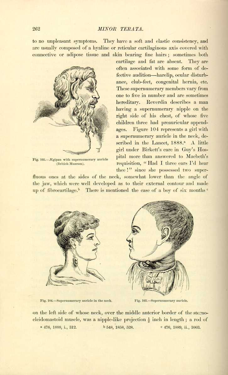
requisition, "Had I three ears I'd hear thee!'' since she possessed two superfluous ones at the sides of the neck, somewhat lower than the angle of the jaw, which were well developed as to their external contour and made up of fibrocartilage. [6.187] There is mentioned the case of a boy of six months [6.188]


Fig. 105.—Supernumerary auricle.
[Description: Drawing of supernumerary auricle]
On the left side of whose neck, over the middle anterior border of the sternocleidomastoid muscle, was a nipple-like projection 1/2 inch in length; a rod of
In some people there is an excessive development of the auricular muscles, enabling them to move their ears in a manner similar to that of the lower animals. Of the celebrated instances the Abbé de Marolles, says Vigneul-Marville, bears witness in his "Mémoires'' that the Regent Crassot could easily move his ears. Saint Augustine mentions this anomaly.
Double tympanitic membrane is spoken of by Loeseke. *[489] There is sometimes natural perforation of the tympanum in an otherwise perfect ear, which explains how some people can blow tobacco-smoke from the ear. Fournier [6.190] has seen several Spaniards and Germans who could perform this feat, and knew one man who could smoke a whole cigar without losing any smoke, since he made it leave either by his mouth, his ears, or in both ways. Fournier in the same article mentions that he has seen a woman with ears over four inches long.
Strange to say, there have been reports of cases in which the ossicles were deficient without causing any imperfection of hearing. Caldani [6.191] mentions a case with the incus and malleus deficient, and Scarpa *[715] and Torreau [6.192] quote instances of deficient ossicles. Thomka in 1895 reported a case of supernumerary tympanic ossicle, the nature of which was unknown, although it was neither an inflammatory product nor a remnant of Meckel's cartilage.
Absence of the Limbs.—Those persons born without limbs are either the subjects of intrauterine amputation or of embryonic malformation. Probably the most celebrated of this class was Marc Cazotte, otherwise known as "Pepin,'' who died in Paris in the last century at the age of sixty-two of a chronic intestinal disorder. He had no arms, legs, or scrotum, but from very jutting shoulders on each side were well-formed hands. His abdomen ended in a flattened buttock with badly-formed feet attached. He was exhibited before the public and was celebrated for his dexterity. He performed nearly all the necessary actions, exhibited skilfulness in all his movements, and was credited with the ability of coitus. He was quite intellectual, being able to write in several languages. His skeleton is preserved in the Musée Dupuytren (Fig. 106). Flachsland [6.193] speaks of a woman who three times had borne children without arms and legs. Hastings [6.194] describes a living child born without any traces of arms or legs (Fig. 107). Garlick [6.195] has seen a child with neither upper nor lower extremities. In place of them were short stumps three or four inches long, closely resembling the ordinary stumps after
Hulke [6.198] describes a child of four whose upper limbs were absent, a small dimple only

Fig. 106.—"Pepin'' (Musée Dupuytren).
[Description: Drawing of "Pepin"]
being in their place. He had free movement of the shoulders in every direction. and could grasp objects between his cheeks and his acromian process; the prehensile power of the toes was well developed,
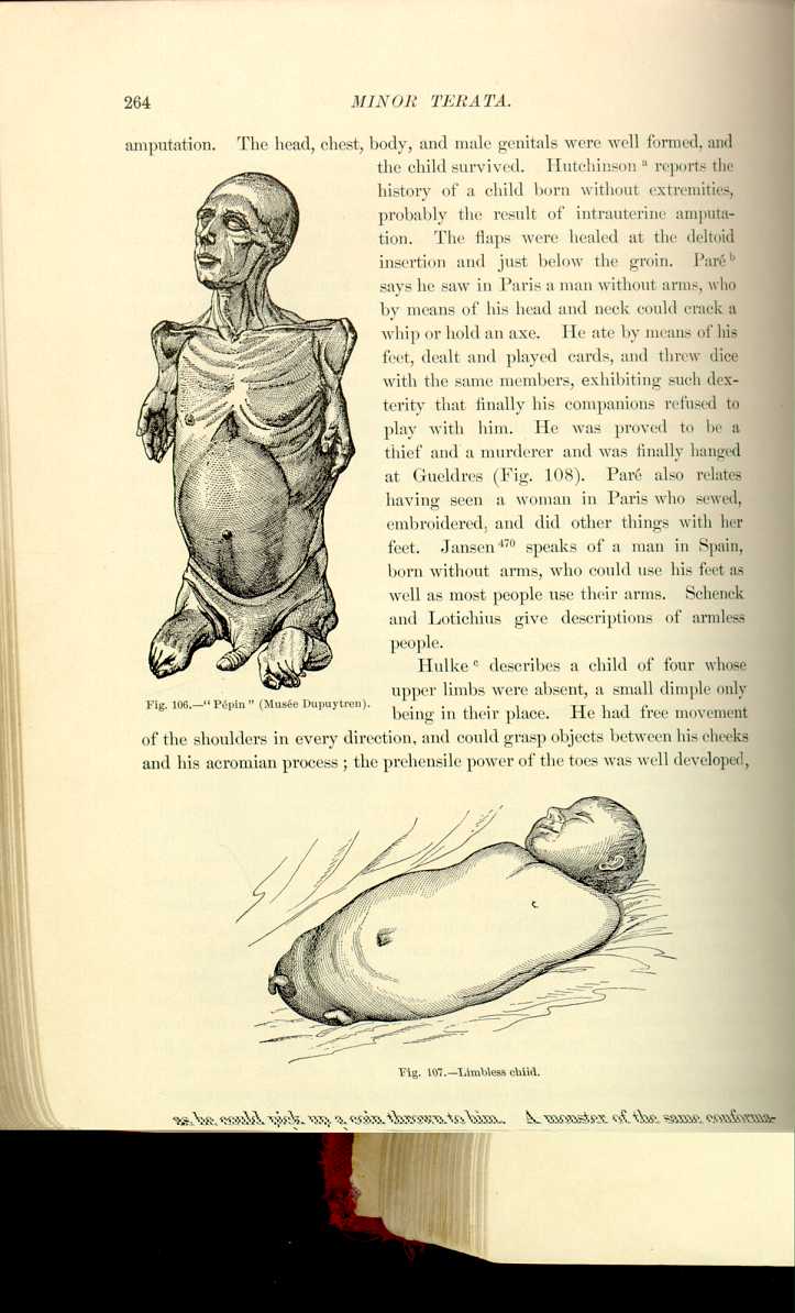
Fig. 107.—Limbless child.
[Description: Drawing of limbless child]
as he could pick up a coin thrown to him. A monster of the same conformation
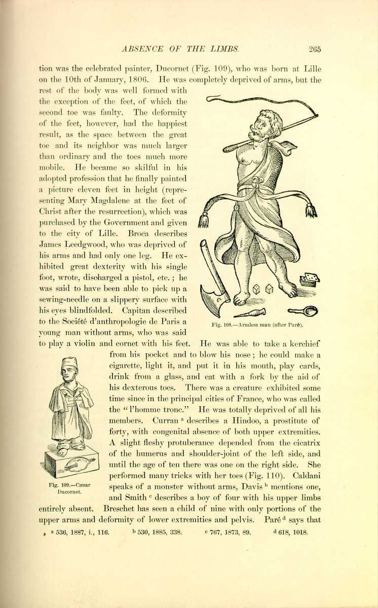
Fig. 108.—Armless man (after Paré).
[Description: Drawing of an armless man]
young man without arms, who was said to play a violin and cornet with his feet. He was able to take a kerchief from his pocket and to blow his nose; he could make a cigarette, light it, and put it in his mouth, play cards, drink from a glass, and eat with a fork by the aid of his dexterous toes. There was a creature exhibited some time since in the principal cities of France, who was called the "l'homme tronc.'' He was totally deprived of all his members. Curran [6.199] describes a Hindoo, a prostitute of forty, with congenital absence of both upper extremities. A slight fleshy protuberance depended from the cicatrix of the humerus and shoulder-joint of the left side, and until the age of ten there was one on the right side. She performed many tricks with her toes (Fig.110). Caldani

Fig. 109.—Cæsar
Ducornet.
[Description: Drawing of Cæsar Ducornet]
speaks of a monster without arms, Davis [6.200] mentions one, and Smith [6.201] describes a boy of four with his upper limbs entirely absent. Breschet has seen a child of nine with only portions of the upper arms and deformity of lower extremities and pelvis. Paré [6.202] says that
The figure and skeleton of Harvey Leach, called "Hervio Nono,'' is in the museum of the University College in London. The pelvis was comparatively weak, the femurs hardly to be recognized, and the right tibia and foot defective; the left foot was better developed, although far from being in due proportion to the trunk above. He was one of the most remarkable gymnasts of his day, and notwithstanding the distortion of his lower limbs had marvelous power and agility in them. As an arena-horseman, either standing or sitting, he was scarcely excelled. He walked and even ran quite well, and his power of leaping, partly with his feet and partly

Fig. 110.—Hindoo armless
woman (Curran).
[Description: Drawing of a Hindoo armless woman]
with his hands, was unusual. His lower limbs were so short that, erect, he touched the floor with his fingers, but he earned his livelihood as much with his lower as with his upper limbs. In his skeleton his left lower limb, between the hip and heel, measured 16 inches, while the right, between the same points, measured nine inches (Fig. 111). [6.203] Hare [6.204] mentions a boy of five and a half whose head and trunk were the same as in any other child of like age. He was 22 1/2 inches high, had no spinal curvature, but was absolutely devoid of lower extremities. The right arm was two inches long and the left 2 1/4 . Each contained the head and a small adjoining portion of the humerus. The legs were represented by masses of cellular tissue and fat covered by skin which projected about an inch. He was intelligent, had a good memory, and exhibited considerable activity. He seemed to have had more than usual mobility and power of flexion of the lower lumbar region. When on his back he was unable to rise up, but resting on the lower part of the pelvis he
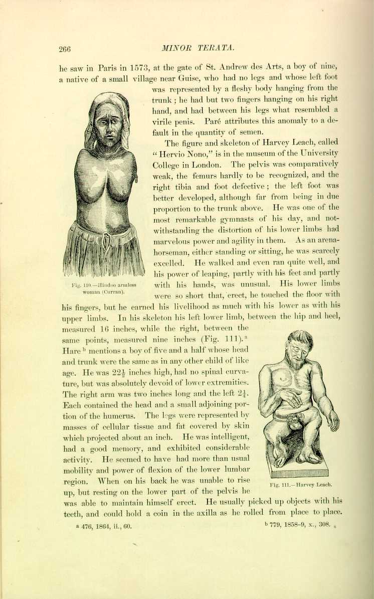
Fig. 111.—Harvey Leach.
[Description: Drawing of Harvey Leach]
was able to maintain himself erect. He usually picked up objects with his teeth, and could hold a coin in the axilla as he rolled from place to place.
Parvin [6.205] describes a "turtle-man'' as an ectromelian, almost entering the class of phocomelians or seal-like monsters; the former term signifies abortive or imperfect formation of the members. The hands and feet were normally developed, but the arms, forearms, and legs are much shortened (see page 84.).
The "turtle-woman'' of Demerara [6.206] (Fig. 114) was so called because her mother when pregnant was frightened by a turtle, and also from the child's fancied resemblance to a turtle. The femur was six inches long, the woman had a foot of six bones, four being toes, viz., the first and second phalanges of the first and second toes. She had an acetabulum, capsule, and ligamentum teres, but no tibia or fibula; she also had a defective
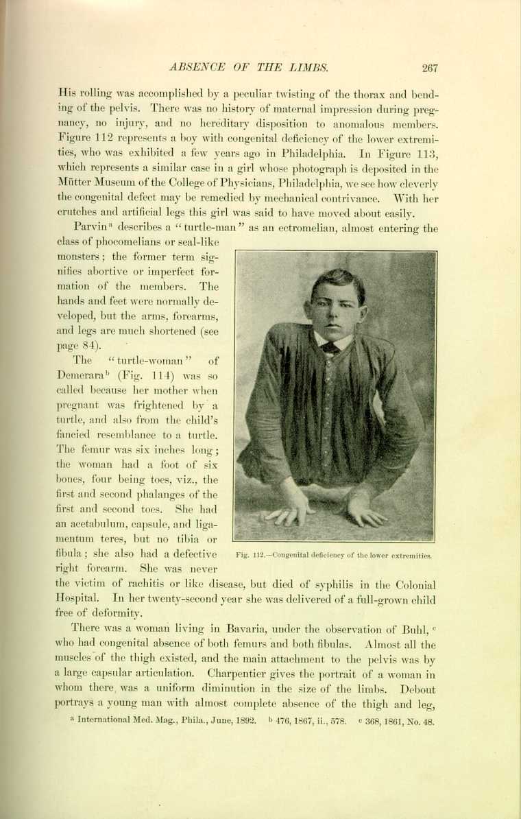
right forearm. She was never the victim of rachitis or like disease, but died of syphilis in the Colonial Hospital. In her twenty-second year she was delivered of a full-grown child free of deformity.
There was a woman living in Bavaria, under the observation of Buhl, [6.207] who had congenital absence of both femurs and both fibulas. Almost all the muscles of the thigh existed, and the main attachment to the pelvis was by a large capsular articulation. Charpentier gives the portrait of a woman in whom there was a uniform diminution in the size of the limbs. Debout portrays a young man with almost complete absence of the thigh and leg,

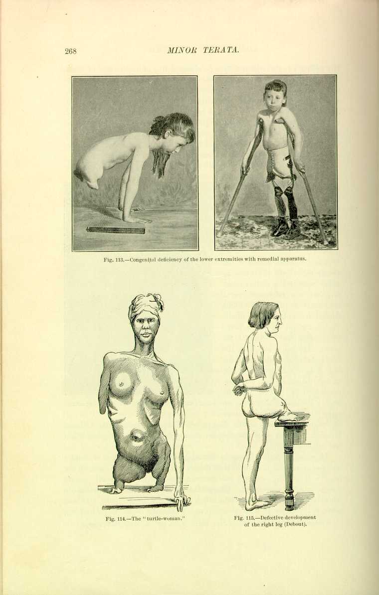
Fig. 114.—The "turtle-woman.''
[Description: Drawing of the "turtle-woman"]

from whose right hip there depended a foot (Fig. 115). Accrell [6.208] describes a peasant of twenty-six, born without a hip, thigh, or leg on the right side. The external genital organs were in their usual place, but there was only one testicle in the scrotum. The man was virile. The rectum instead of opening outward and underneath was deflected to the right.
Supernumerary Limbs.—Haller reports several cases of supernumerary extremities. Plancus [6.209] speaks of an infant with a complete third leg, and Dumeril [6.210] cites a similar instance. Geoffroy-Saint-Hilaire presented to the Académie des Sciences in 1830 a child with four legs and feet who was in good health. Amman saw a girl with a large thigh attached to her nates. Below the thigh was a single leg made by the fusion of two legs. No patella was found and the knee was anchylosed. One of the feet of the supernumerary

Fig. 116.—Gustav Evrard (after Guérin).
[Description: Drawing of Gustav Evrard]
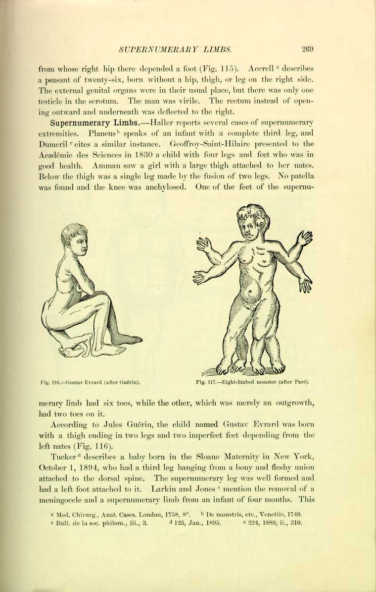
limb had six toes, while the other, which was merely an outgrowth, had two toes on it.
According to Jules Guérin, the child named Gustav Evrard was born with a thigh ending in two legs and two imperfect feet depending from the left nates (Fig. 116).
Tucker [6.211] describes a baby born in the Sloane Maternity in New York, October 1, 1894, who had a third leg hanging from a bony and fleshy union attached to the dorsal spine. The supernumerary leg was well formed and had a left foot attached to it. Larkin and Jones [6.212] mention the removal of a meningocele and a supernumerary limb from an infant of four months. This
Paré [6.213] says that on the day the Venetians and the Genevois made peace a monster was born in Italy which had four legs of equal proportions, and besides had two supernumerary arms from the elbows of the normal limbs. This creature lived and was baptized (Fig. 117).
Anomalies of the Feet.—Hatte [6.214] has seen a woman who bore a child that had three feet. Bull [6.215] gives a description of a female infant with the left foot double or cloven. There was only one heel, but the anterior portion consisted of an anterior and a posterior part. The anterior foot presented a great toe and four smaller ones, but deformed like an example of talipes equinovarus. Continuous with the outer edge of the anterior part and curving beneath it was a posterior part, looking not unlike a second foot, containing

Fig. 118—Double foot (Bull).
[Description: Drawing of a double foot]
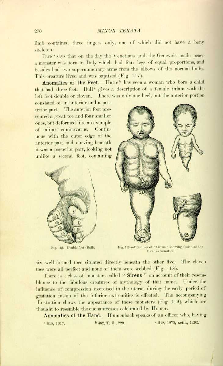
six well-formed toes situated directly beneath the other five. The eleven toes were all perfect and none of them were webbed (Fig. 118).
There is a class of monsters called "Sirens'' on account of their resemblance to the fabulous creatures of mythology of that name. Under the influence of compression exercised in the uterus during the early period of gestation fusion of the inferior extremities is effected. The accompanying illustration shows the appearance of these monsters (Fig. 119), which are thought to resemble the enchantresses celebrated by Homer.
Anomalies of the Hand.—Blumenbach speaks of an officer who, having

Fig.120.—Double hand (Murray).
[Description: Drawing of three views of a double hand]
could be extended while the other seven were flexed (Fig. 120). Giraldès saw an infant in 1864 with somewhat the same deformity, but in which the disposition of the muscles and tendons permitted the ordinary movements (Fig. 121).
Absence of Digits.—Maygrier [6.217] describes a woman of twenty-four who instead of having a hand on each arm had only one finger, and each foot had but two toes. She was delivered of two female children in 1827 and one in 1829, each having exactly the same deformities. Her mother was perfectly formed, but the father had but one toe on his foot and one finger on his left hand.
Köhler [6.218] gives photographs of quite a remarkable case
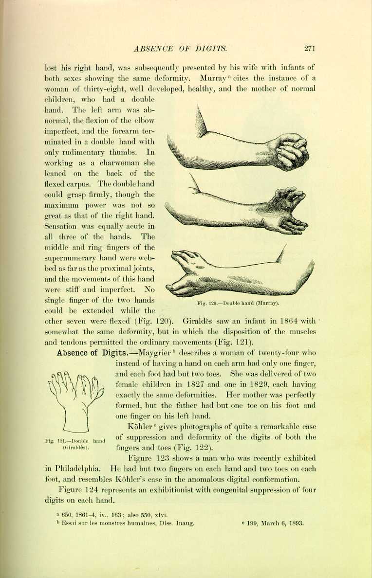
Fig, 121.—Double hand
(Giraldès).
[Description: Drawing of a double hand]
of suppression and deformity of the digits of both the fingers and toes (Fig. 122).
Figure 123 shows a man who was recently exhibited in Philadelphia. He had but two fingers on each hand and two toes on each foot, and resembles Köhler's case in the anomalous digital conformation.
Figure 124 represents an exhibitionist with congenital suppression of four digits on each hand.
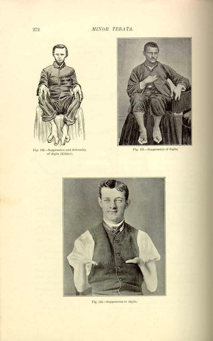

Fig. 123.—Suppression of digits.
[Description: Photograph of man whose digits are suppressed]

Tubby [6.219] has seen a boy of three in whom the first, second, and third toes of each foot were suppressed, the great toe and the little toe being so overgrown that they could be opposed. In this family for four generations 15 individuals out of 22 presented this defect of the lower extremity. The patient's brothers and a sister had exactly the same deformity, which has been called "lobster-claw foot.''
Falla of Jedburgh speaks of an infant who was born without forearms or hands; at the elbow there was a single finger attached by a thin string of tissue. This was the sixth child, and it presented no other deformity. Falla also says that instances of intrauterine digital amputation are occasionally seen.
According to Annandale, supernumerary digits may be classified as follows:—
(1) A deficient organ, loosely attached by a narrow pedicle to the hand or foot (or to another digit).
(2) A more or less developed organ, free at its extremity, and articulating with the head or sides of a metacarpal, metatarsal, or phalangeal bone.
(3) A fully developed separate digit.
(4) A digit intimately united along its whole length with another digit, and having either an additional metacarpal or metatarsal bone of its own, or articulating with the head of one which is common to it and another digit.
Superstitions relative to supernumerary fingers have long been prevalent. In the days of the ancient Chaldeans it was for those of royal birth especially that divinations relative to extra digits were cast. Among the ancients we also occasionally see illustrations emblematic of wisdom in an individual with many fingers, or rather double hands, on each arm.
Hutchinson, [6.220] in his comments on a short-limbed, polydactylous dwarf (Fig. 125) which was dissected by Ruysch, the celebrated Amsterdam anatomist, writes as follows.—
"This quaint figure is copied from Theodore Kerckring's `Spicilegium Anatomicum,' published in Amsterdam in 1670. The description states that the body was that of an infant found drowned in the river on October 16, 1668. It was dissected by the renowned Ruysch. A detailed description of the skeleton is given. My reason for now reproducing the plate is that it offers an important item of evidence in reference to the development of short-limbed dwarfs. Although we must not place too much reliance on the accuracy of the draughtsman, since he has figured some superfluous lumbar vertebræ, yet there can be no doubt that the limbs are much too short for the trunk and head. This remark especially applies to the lower limbs and pelvis. These are exactly like those of the Norwich dwarf and of the skeleton in the Heidelberg Museum which I described in a recent number of the `Archives.' The point of extreme interest in the present case
"This occurrence of short limbs with polydactylism seems to prove conclusively that the condition may be due to a modification of development of a totally different nature from rickets. It is probable that the infant was not at full term. Among the points which the author has noticed in his description are that the fontanelle was double its usual size; that the orbits were somewhat deformed; that the two halves of the lower jaw were already united; and that the ribs were short and badly formed. He also, of course, draws attention to the shortness of the limbs, the stoutness of the long bones, and the supernumerary digits. I find no statement that the skeleton was
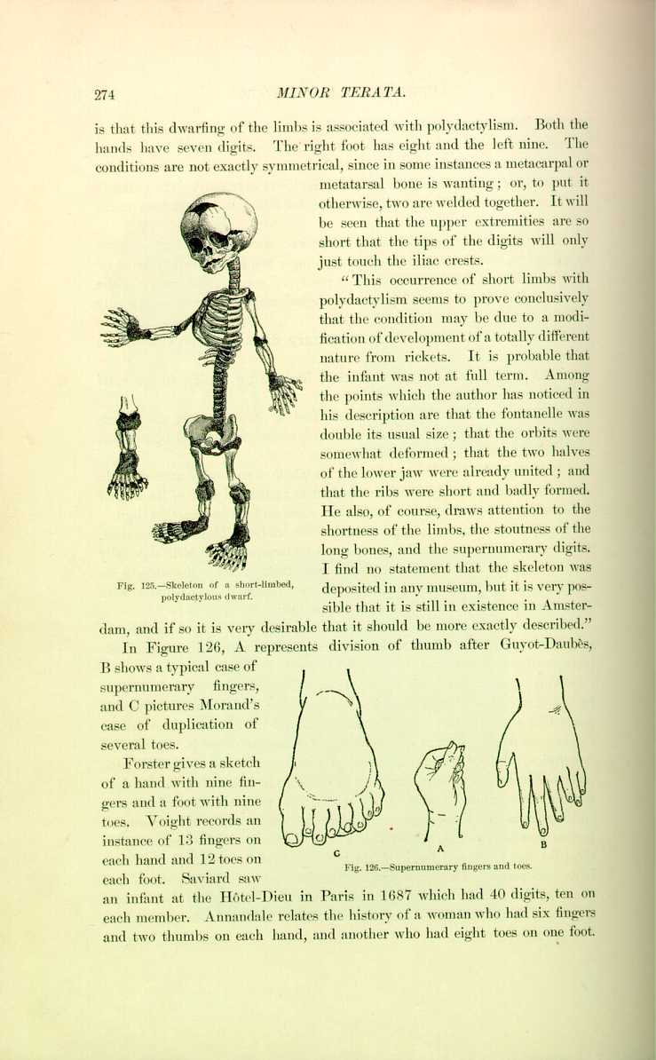
deposited in any museum, but it is very possible that it is still in existence in Amsterdam, and if so it is very desirable that it should be more exactly described,''
In Figure 126, A represents division of thumb after Guyot-Daubès, shows a typical case of supernumerary fingers, and C pictures Morand's case of duplication of several toes.
Forster gives a sketch of a hand with nine fingers and a foot with nine toes. Voight records an instance of 13 fingers on each hand and 12 toes on

Fig. 126.—Supernumerary fingers and toes.
[Description: Drawing of supernumerary fingers and toes]
each foot. Saviard saw an infant at the Hôtel-Dieu in Paris in 1687 which had 40 digits, ten on each member. Annandale relates the history of a woman who had six fingers and two thumbs on each hand, and another who had eight toes on one foot.
The influence of heredity on this anomaly is well demonstrated. Réaumur was one of the first to prove this, as shown by the Kelleia family of Malta, and there have been many corroboratory instances reported; it is shown to last for three, four, and even five generations; intermarriage with normal persons finally eradicates it.
It is particularly in places where consanguineous marriages are prevalent that supernumerary digits persist in a family. The family of Foldi in the tribe of Hyabites living in Arabia are very numerous and confine their marriages to their tribe. They all have 24 digits, and infants born with the normal number are sacrificed as being the offspring of adultery. The inhabitants of the village of Eycaux in France, at the end of the last century, had nearly all supernumerary digits either on the hands or feet. Being isolated in an inaccessible and mountainous region, they had for many years intermarried and thus perpetuated the anomaly. Communication being opened, they emigrated or married strangers and the sexdigitism vanished. Maupertuis recalls the history of a family living in Berlin whose members had 24 digits for many generations. One of them being presented with a normal infant refused to acknowledge it. There is an instance in the Western United States [6.226] in which supernumerary digits have lasted through five generations. Cameron [6.227] speaks of two children in the same family who were polydactylic, though not having the same number of supernumerary fingers.
Smith and Norwell [6.228] report the case of a boy of fifteen both of whose hands showed webbing of the middle and ring fingers and accessory nodules of bone between the metacarpals, and six toes on each foot. The boy's father showed similar malformations, and in five generations 21 out of 28 individuals were thus malformed, ten females and 11 males. The deformity was especially transmitted in the female line.
Instances of supernumerary thumbs are cited by Panaroli, [6.229] Ephemerides, Munconys, as well as in numerous journals since. This anomaly is
Hypertrophy of the digits is the result of many different processes, and true hypertrophy or gigantism must be differentiated from acromegaly, elephantiasis, leontiasis, and arthritis deformans, for which distinction the reader is referred to an article by Park. [6.230] Park also calls attention to the difference between acquired gigantism, particularly of the finger and toes, and another condition of congenital gigantism, in which either after or before birth there is a relatively disproportionate, sometimes enormous, overgrowth of perhaps one finger or two, perhaps of a limited portion of a hand or foot, or possibly of a part of one of the limbs. The best collection of this kind of specimens is in the College of Surgeons in London.
Curling [6.231] quotes a most peculiar instance of hypertrophy of the fingers in a sickly girl (Fig. 127). The middle and ring fingers of the right hand were of unusual size, the middle finger measuring 5 1/2 inches in length four inches in circumference. On the left hand the thumb and middle fingers were hypertrophied and the index finger was as long as the middle one of the right hand. The middle finger had a lateral curvature outward, due to a displacement of the extensor tendon. This affection resembled
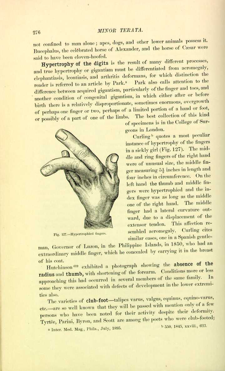
Fig. 127.—Hypertrophied fingers.
[Description: Drawing of a hand with hypertrophied fingers]
acromegaly. Curling cites similar cases, one in a Spanish gentleman, Governor of Luzon, in the Philippine Islands, in 1850, who had an extraordinary middle finger, which he concealed by carrying it in the breast of his coat.
Hutchinson *[619] exhibited a photograph showing the absence of the radius and thumb, with shortening of the forearm. Conditions more or less approaching this had occurred in several members of the same family. In some they were associated with defects of development in the lower extremities also.
The varieties of club-foot—talipes varus, valgus, equinus, equino-varus, etc.—are so well known that they will be passed with mention only of a few persons who have been noted for their activity despite their deformity. Tyrtée, Parini, Byron, and Scott are among the poets who were club-footed;
Transpositions of the character of the vertebræ are sometimes seen. In man the lumbar vertebræ have sometimes assumed the character of the sacral vertebræ, the sacral vertebræ presenting the aspect of lumbar vertebræ, etc. It is quite common to see the first lumbar vertebra presenting certain characteristics of the dorsal.
Numerical anomalies of the vertebræ are quite common, generally in the lumbar and dorsal regions, being quite rare in the cervical, although there have been instances of six or eight cervical vertebræ. In the lower animals the vertebræ are prolonged into a tail, which, however, is sometimes absent, particularly when hereditary influence exists. It has been noticed in the class of dogs whose tails are habitually amputated to improve their appearance that the tail gradually decreases in length. Some breeders deny this fact.
Human Tails.—The prolongation of the coccyx sometimes takes the shape of a caudal extremity in man. Broca and others claim that the sacrum and the coccyx represent the normal tail of man, but examples are not infrequent in which there has been a fleshy or bony tail appended to the coccygeal region. Traditions of tailed men are old and widespread, and tailed races were supposed to reside in almost every country. There was at one time an ancient belief that all Cornishmen had tails, and certain men of Kent were said to have been afflicted with tails in retribution for their insults to Thomas à Becket. Struys, a Dutch traveler in Formosa in the seventeenth century, describes a wild man caught and tied for execution who had a tail more than a foot long, which was covered with red hair like that of a cow.
The Niam Niams of Central Africa are reported to have tails smooth and hairy and from two to ten inches long. Hubsch of Constantinople remarks that both men and women of this tribe have tails. Carpus, or Berengarius Carpensis, as he is called, in one of his Commentaries said that there were some people in Hibernia with long tails, but whether they were fleshy or cartilaginous could not be known, as the people could not be approached. Certain supposed tailed races which have been described by sea-captains and voyagers are really only examples of people who wear artificial appendages about the waists, such as palm-leaves and hair. A certain Wesleyan missionary, George Brown, in 1876 spoke of a formal breeding of a tailed race in Kali, off the coast of New Britain. Tailless children were slain at once, as they would be exposed to public ridicule. The tailed men of Borneo are
There was reported from Caracas [6.232] the discovery of a tribe of Indians in Paraguay who were provided with tails. The narrative reads somewhat after this manner: One day a number of workmen belonging to Tacura Tuyn while engaged in cutting grass had their mules attacked by some Guayacuyan Indians. The workmen pursued the Indians but only succeeded in capturing a boy of eight. He was taken to the house of Señor Francisco Galeochoa at Posedas, and was there discovered to have a tail ten inches long. On interrogation the boy stated that he had a brother who had a tail as long as his own, and that all the tribe had tails.
Aetius, Bartholinus, Falk, Harvey, Kölping, Hesse, Paulinus, Strauss, and Wolff give descriptions of tails. Blanchard *[213] says he saw a tail fully a span in length: and there is a description in 1690 of a man by the name of Emanuel König, a son of a doctor of laws *[570] who had a tail half a span long, which grew directly downward from the coccyx and was coiled on the perineum, causing much discomfort. Jacob [6.233] describes a pouch of skin resembling a tail which hung from the tip of the coccyx to the length of six inches. It was removed and was found to be thicker than the
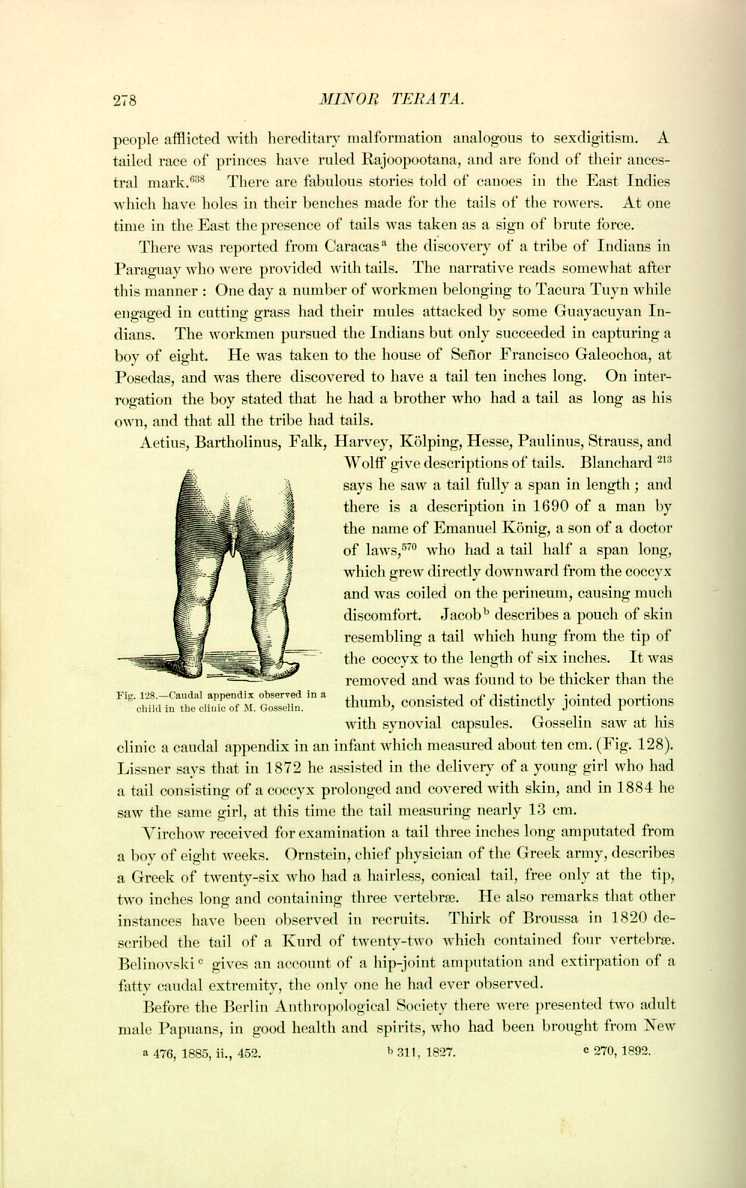
thumb, consisted of distinctly jointed portions with synovial capsules. Gosselin saw at his clinic a caudal appendix in an infant which measured about ten cm. (Fig. 128). Lissner says that in 1872 he assisted in the delivery of a young girl who had a tail consisting of a coccyx prolonged and covered with skin, and in 1884 he saw the same girl, at this time the tail measuring nearly 13 cm.
Virchow received for examination a tail three inches long amputated from a boy of eight weeks. Ornstein, chief physician of the Greek army, describes a Greek of twenty-six who had a hairless, conical tail, free only at the tip, two inches long and containing three vertebræ. He also remarks that other instances have been observed in recruits. Thirk of Broussa in 1820 described the tail of a Kurd of twenty-two which contained four vertebræ. Belinovski [6.234] gives an account of a hip-joint amputation and extirpation of a fatty caudal extremity, the only one he had ever observed.
Before the Berlin Anthropological Society there were presented two adult male Papuans, in good health and spirits, who had been brought from New
Bartels [6.236] presents a very complete article in which he describes 21 persons born with tails, most of the tails being merely fleshy protuberances. Darwin *[289] speaks of a person with a fleshy tail and refers to a French article on human tails. [6.237]
Science [6.238] contains a description of a negro child born near Louisville, eight weeks old, with a pedunculated tail 2 1/2 inches long, with a base 1 1/4 inches in circumference. The tail resembled in shape a pig's tail and had grown 1/4 inch since birth. It showed no signs of cartilage or bone, and had its origin from a point slightly to the left of the median line and about an inch above the end of the spinal column.
Dickinson [6.239] recently reported the birth of a child with a tail (Fig. 129). It was a well-developed female between 5 1/2 and six pounds in weight. The coccyx was covered with the skin on both the anterior and posterior surfaces. It thus formed a tail of the size of the nail of the little finger, with a length of nearly 3/16 inch on the inner surface and 3/8 inch on the rear surface. This little tip could be raised from the body and it slowly sank back.
In addition to the familiar caudal projection of the human fetus, Dickinson mentions a group of other vestigial remains of a former state of things. Briefly these are:—
(1) The plica semilunaris as a vestige of the nictitating membrane of certain birds.
(2) The pointed ear, or the turned-down tip of the ears of many men.
(3) The atrophied muscles, such as those that move the ear, that are well developed in certain people, or that shift the scalp, resembling the action of a horse in ridding itself of flies.
(4) The supracondyloid foremen of the humerus.
(5) The vermiform appendix.
(6) The location and direction of the hair on the trunk and limbs.
(7) The dwindling wisdom-teeth.
(8) The feet of the fetus strongly deflected inward, as in the apes, and persisting in the early months of life, together with great mobility and a distinct projection of the great toe at an angle from the side of the foot.
Horrocks [6.240] ascribes to these anal tags a pathologic importance. He claims that they may be productive of fistula in ano, superficial ulcerations, fecal concretions, fissure in ano, and that they may hypertrophy and set up tenesmus and other troubles. The presence of human tails has given rise to discussion between friends and opponents of the Darwinian theory. By some it is
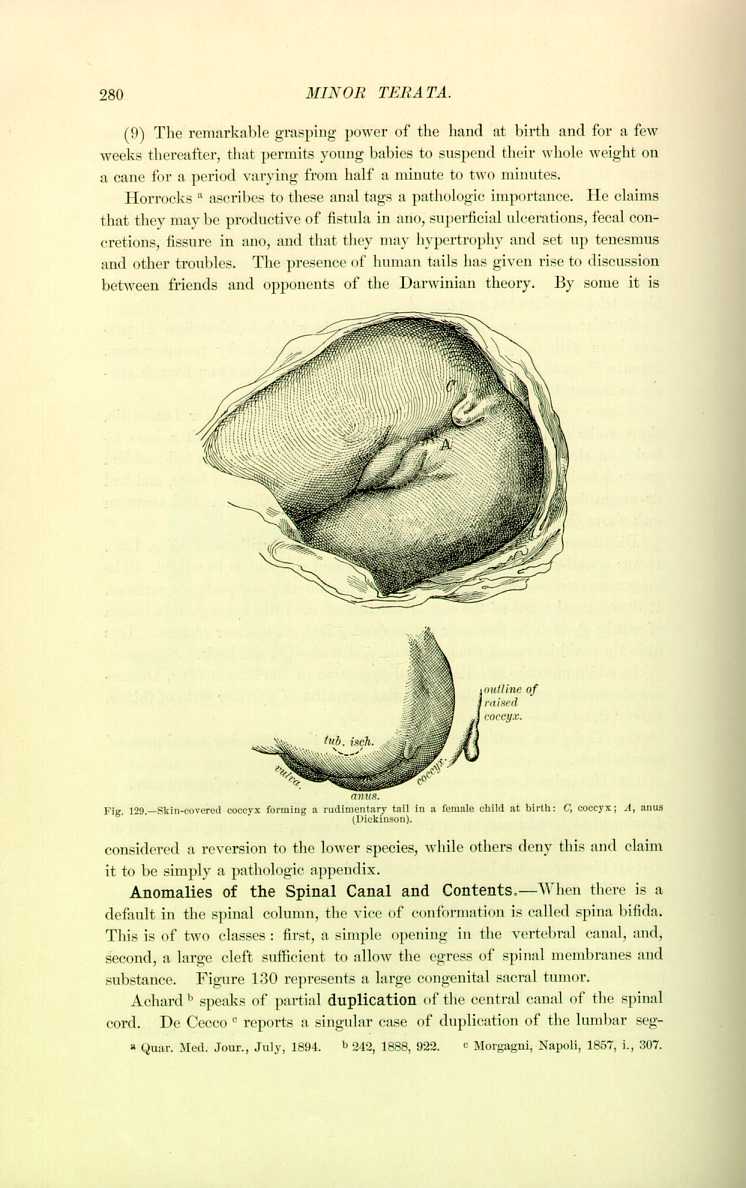
considered a reversion to the lower species, while others deny this and claim it to be simply a pathologic appendix.
Anomalies of the Spinal Canal and Contents.—When there is a default in the spinal column, the vice of conformation is called spina bifida. This is of two classes: first, a simple opening in the vertebral canal, and, second, a large cleft sufficient to allow the egress of spinal membranes and substance. Figure 130 represents a large congenital sacral tumor.
Achard [6.241] speaks of partial duplication of the central canal of the spinal cord. De Cecco [6.242] reports a singular case of duplication of the lumbar segment
Foot [6.243] records a case of amyelia, or absence of the spinal cord, in a fetus with hernia cerebri and complete fissure of the spinal column. Nicoll and Arnold [6.244] describe an anencephalous fetus with absence of spinal marrow; and Smith also records the birth of an amyelitic fetus. [6.245]
In some persons there are exaggerated curvatures of the spine. The first of these curvatures is called kyphosis, in which the curvature is posterior; second, lordosis, in which the curvature is anterior; third, scoliosis, in which it is lateral, to the right or left.
Kyphosis is the most common of the deviations in man and is most often found in the dorsal region, although it may be in the lumbar region. Congenital kyphosis is very

rare in man, is generally seen in monsters, and when it does exist is usually accompanied by lordosis or spine bifida. We sometimes observe a condition of anterior curvature of the lumbar and sacral regions, which might be taken for a congenital lordosis, but this is really a deformity produced after birth by the physiologic weight of the body. Figure 131 represents a case of lordosis caused by paralysis of the spinal muscles.
Analogous to this is what the accoucheurs call spondylolisthesis. Scoliosis may be a cervicodorsal, dorsolumbar, or lumbosacral curve, and the inclination of the vertebral column may be to the right or left (Figs. 132 and 133). The pathologists divide scoliosis into a myopathic variety, in which the trouble is a physiologic antagonism of the muscles; or osteopathic, ordinarily associated with rachitis, which latter variety is generally accountable for congenital scoliosis. In some cases the diameter of the chest is shortened to an almost incredible degree, but may yet be compatible with life. Glover [6.246] speaks of an extraordinary deformity of the chest with lateral curvature of the spine, in which the diameter from the pit of the stomach to the spinal integument was only 5 1/2 inches.
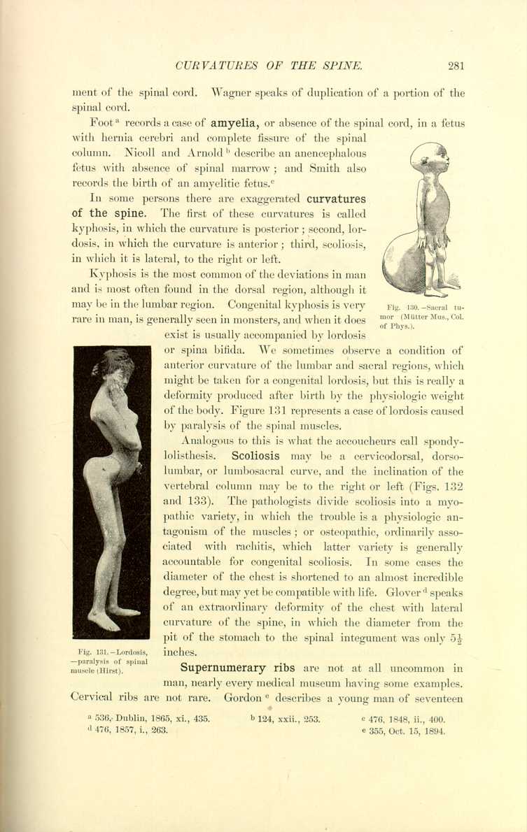
Supernumerary ribs are not at all uncommon in man, nearly every medical museum having some examples. Cervical ribs are not rare. Gordon [6.247] describes a young man of seventeen
In some cases we see fissure of the sternum, caused either by deficient union or absence of one of its constituent parts. In the most exaggerated cases these fissures permit the exit of the heart, and as a general rule ectopies
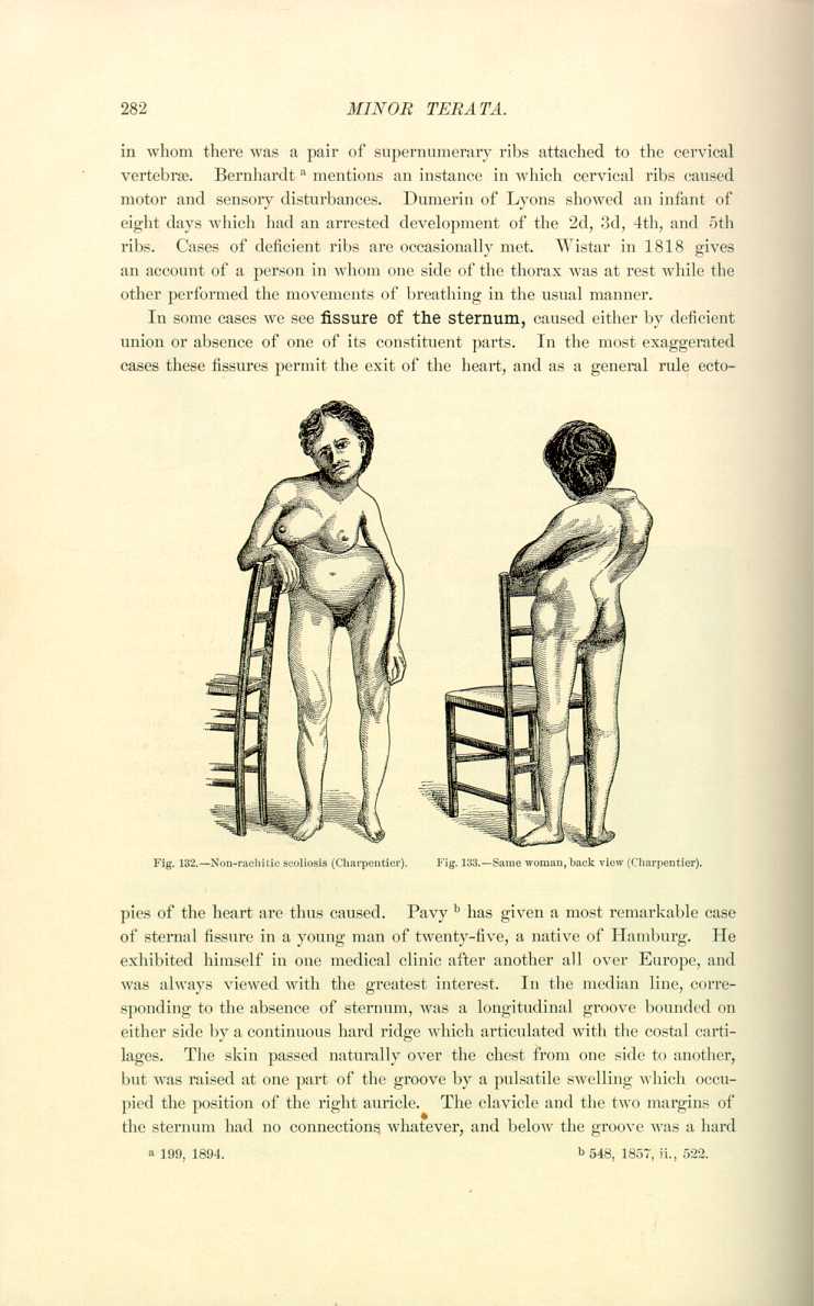
of the heart are thus caused. Pavy [6.249] has given a most remarkable case of sternal fissure in a young man of twenty-five, a native of Hamburg. He exhibited himself in one medical clinic after another all over Europe, and was always viewed with the greatest interest. In the median line, corresponding to the absence of sternum, was a longitudinal groove bounded on either side by a continuous hard ridge which articulated with the costal cartilages. The skin passed naturally over the chest from one side to another, but was raised at one part of the groove by a pulsatile swelling which occupied the position of the right auricle. The clavicle and the two margins of the sternum had no connections whatever, and below the groove was a hard
Madden [6.250] pictures (Figs. 134 and 135) a Swede of forty with congenital
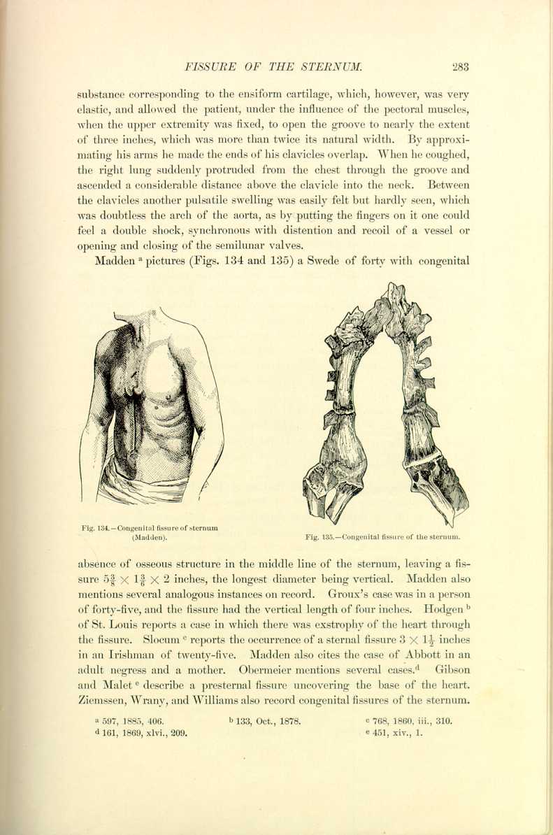

absence of osseous structure in the middle line of the sternum, leaving a fissure 5 3/8 X 1 3/6 X 2 inches, the longest diameter being vertical. Madden also mentions several analogous instances on record. Groux's case was in a person of forty-five, and the fissure had the vertical length of four inches. Hodgen [6.251] of St. Louis reports a case in which there was exstrophy of the heart through the fissure. Slocum [6.252] reports the occurrence of a sternal fissure 3 X 1 1/2 inches in an Irishman of twenty-five. Madden also cites the case of Abbott in an adult negress and a mother. Obermeier mentions several cases. [6.253] Gibson and Malet [6.254] describe a presternal fissure uncovering the base of the heart. Ziemssen, Wrany, and Williams also record congenital fissures of the sternum.
Some preliminary knowledge of embryology is essential to understand the formation of branchial fissures, and we refer the reader to any of the standard works on embryology for this information. Dzondi was one of the first to recognize and classify congenital fistulas of the neck. The proper classification is into lateral and median fissures. In a case studied by Février [6.257] the exploration of a lateral pharyngeal fistula produced by the introduction of the sound violent reflex phenomena, such as pallor of the face and irregular, violent beating of the heart. The rarest of the lateral class is the preauricular fissure, which has been observed by Février, Le Dentu, Marchand, peyrot, and Routier.
The median congenital fissures of the neck are probably caused by defective union of the branchial arches, although Arndt thinks that he sees in these median fistulas a persistence of the hypobranchial furrow which exists normally in the amphioxus. They are less frequent than the preceding variety.
The most typical form of malformation of the esophagus is imperforation or obliteration. Van Cuyck of Brussels in 1824 delivered a child which died on the third day from malnutrition. Postmortem it was found that the inferior extremity of the esophagus to the extent of about two inches was converted into a ligamentous cord. Porro [6.258] describes a case of congenital obliteration of the esophagus which ended in a cecal pouch about one inch below the inferior portion of the glottidean aperture and from this point to the stomach only measured an inch; there was also tracheal communication. The child was noticed to take to the breast with avidity, but after a little suckling it would cough, become livid, and reject most of the milk through the nose, in this way almost suffocating at each paroxysm; it died on the third day.
In some cases the esophagus is divided, one portion opening into the bronchial or other thoracio organs. Brentano [6.259] describes an infant dying ten days after birth whose esophagus was divided into two portions, one
Anomalies of the Lungs.—Carper describes a fetus of thirty-seven weeks in whose thorax he found a very voluminous thymus gland but no lungs. These organs were simply represented by two little oval bodies having no lobes, with the color of the tissue of the liver. The heart had only one cavity but all the other organs were perfectly formed. This case seems to be unique. Tichomiroff [6.260] records the case of a woman of twenty-four who died of pneumonia in whom the left lung was entirely missing. No traces of a left bronchus existed. The subject was very poorly developed physically. Tichomiroff finds four other cases in literature, in all of which the left lung was absent. Théremin and Tyson record cases of the absence of the left lung.
Supplementary pulmonary lobes are occasionally seen in man and are taken by some authorities to be examples of retrogressive anomalies tending to prove that the derivation of the human race is from the quadrupeds which show analogous pulmonary malformation. Eckley [6.261] reports an instance of
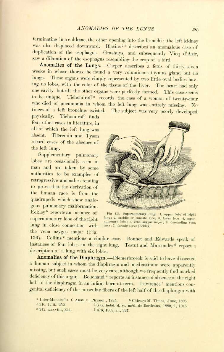
supernumerary lobe of the right lung in close connection with the vena azygos major (Fig. 136). Collins [6.262] mentions a similar case. Bonnet and Edwards speak of instances of four lobes in the right lung. Testut and Marcondès [6.263] report a description of a lung with six lobes.
Anomalies of the Diaphragm.—Diemerbroeck is said to have dissected a human subject in whom the diaphragm and mediastinum were apparently missing, but such cases must be very rare, although we frequently find marked deficiency of this organ. Bouchand [6.264] reports an instance of absence of the right half of the diaphragm in an infant born at term. Lawrence [6.265] mentions congenital deficiency of the muscular fibers of the left half of the diaphragm with
Anomalies of the Stomach.—The Ephemerides contains the account of a dissection in which the stomach was found wanting, and also speaks of two instances of duplex stomach. Bartholinus, *[190] Heister, Hufeland, Morgagni, Riolan, and Sandifort [6.275] cite examples of duplex stomach. Bonet speaks of a case of vomiting which was caused by a double stomach. Struthers [6.276] reports two cases in which there were two cavities to the stomach. Struthers also mentions that Morgagni, Home, Monro, Palmer, Larry, Blasius, Hufeland, and Walther also record instances in which there was contraction in the
Hernia of the stomach is not uncommon, especially in diaphragmatic or umbilical deficiency. There are many cases on record, some terminating fatally from strangulation or exposure to traumatism. Paterson [6.280] reports a case of congenital hernia of the stomach into the left portion of the thoracic cavity. It was covered with fat and occupied the whole left half of the thoracic cavity. The spleen, pancreas, and transverse colon were also superior to the diaphragm. Death was caused by a well-defined round perforation at the cardiac curvature the size of a sixpence.
Anomalies of the Intestines.—The Ephemerides contains the account of an example of double cecum, and Alexander [6.281] speaks of a double colon, and there are other cases of duplication of the bowel recorded. There is an instance of coalition of the jejunum with the liver, *[104] and Treuner [6.282] parallels this case. Aubery, Charrier Poelman,
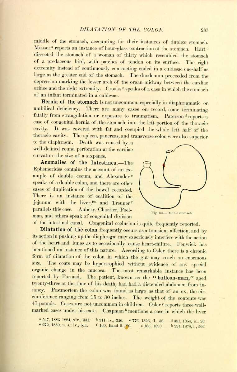
Fig. 137. -Double stomach.
[Description: Illustration of a double stomach]
and others speak of congenital division of the intestinal canal. Congenital occlusion is quite frequently reported.
Dilatation of the colon frequently occurs as a transient affection, and by its action in pushing up the diaphragm may so seriously interfere with the action of the heart and lungs as to occasionally cause heart-failure. Fenwick has mentioned an instance of this nature. According to Osler there is a chronic form of dilatation of the colon in which the gut may reach an enormous size. The coats may be hypertrophied without evidence of any special organic change in the mucosa. The most remarkable instance has been reported by Formad. The patient, known as the "balloon-man,'' aged twenty-three at the time of his death, had had a distended abdomen from infancy. Postmortem the colon was found as large as that of an ox, the circumference ranging from 15 to 30 inches. The weight of the contents was 47 pounds. Cases are not uncommon in children. Osler [6.283] reports three well-marked cases under his care. Chapman [6.284] mentions a case in which the liver
Imperforate Anus.—Cases in which the anus is imperforate or the rectum ends in a blind pouch are occasionally seen. In some instances the rectum is entirely absent, the colon being the termination of the intestinal tract. There are cases on record in which the rectum communicated with the anus solely by a fibromuscular cord. Anorectal atresia is the ordinary imperforation of the anus, in which the rectum terminates in the middle of the sacral cavity. The rectum may be deficient from the superior third of the sacrum, and in this position is quite inaccessible for operation.
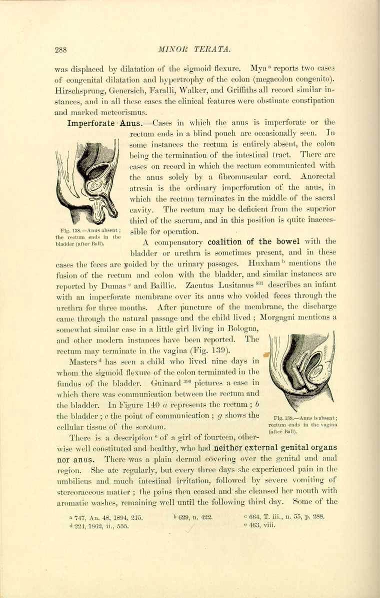
A compensatory coalition of the bowel with the bladder or urethra is sometimes present, and in these cases the feces are voided by the urinary passages. Huxham [6.286] mentions the fusion of the rectum and colon with the bladder, and similar instances are reported by Dumas [6.287] and Baillie. Zacutus Lusitanus *[831] describes an infant with an imperforate membrane over its anus who voided feces through the urethra for three months. After puncture of the membrane, the discharge came through the natural passage and the child lived; Morgagni mentions a somewhat similar case in a little girl living in Bologna, and other modern instances have been reported. The rectum may terminate in the vagina (Fig. 139). Masters [6.288] has seen a child who lived nine days in whom the sigmoid flexure of the colon terminated in the fundus of the bladder. Guinard *[390] pictures a case in which there was communication between the rectum and the bladder. In Figure 140 a represents the rectum; b the bladder; c the point of communication; g shows the

cellular tissue of the scrotum.
There is a description [6.289] of a girl of fourteen, otherwise well constituted and healthy, who had neither external genital organs nor anus. There was a plain dermal covering over the genital and anal region. She ate regularly, but every three days she experienced pain in the umbilicus and much intestinal irritation, followed by severe vomiting of stercoraceous matter; the pains then ceased and she cleansed her mouth with aromatic washes, remaining well until the following third day. Some of the
Fournier [6.290] narrates that he was called by three students, who had been trying to deliver a woman for five days. He found a well-constituted woman of twenty-two in horrible agony, who they said had not had a passage of the bowels for eight days, so he prescribed an enema. The student who was directed to give the enema found to his surprise that there was no anus, but by putting his finger in the vagina he could discern the floating end of the rectum, which was full of feces. There was an opening in this suspended rectum about the size of an undistended anus. Lavage was practiced by a cannula introduced through the opening, and a great number of cherry stones
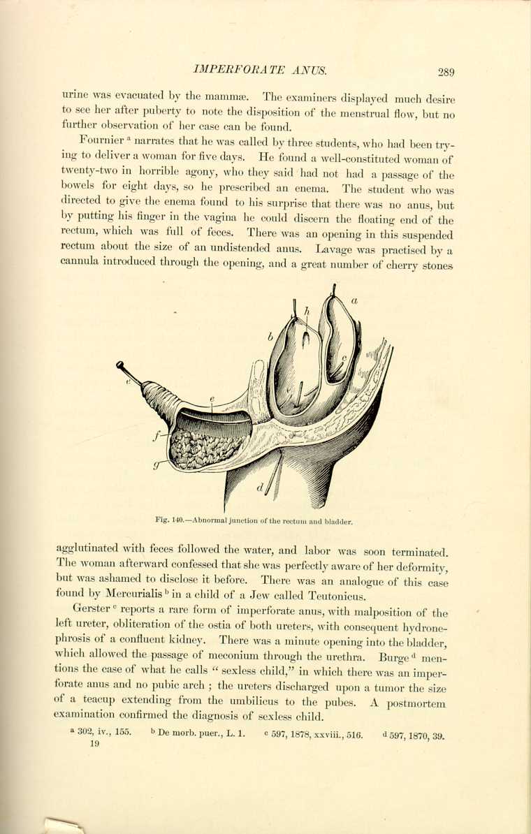
agglutinated with feces followed the water, and labor was soon terminated. The woman afterward confessed that she was perfectly aware of her deformity, but was ashamed to disclose it before. There was an analogue of this case found by Mercurialis [6.291] in a child of a Jew called Teutonicus.
Gerster [6.292] reports a rare form of imperforate anus, with malposition of the left ureter, obliteration of the ostia of both ureters, with consequent hydronephrosis of a confluent kidney. There was a minute opening into the bladder, which allowed the passage of meconium through the urethra. Burge [6.293] mentions the case of what he calls "sexless child,'' in which there was an imperforate anus and no pubic arch; the ureters discharged upon a tumor the size of a teacup extending from the umbilicus to the pubes. A postmortem examination confirmed the diagnosis of sexless child.
Laget [6.298] reports an instance of supernumerary lobe in the liver. Van Buren [6.299] describes a supernumerary liver. Sometimes there is rotation, real or apparent, caused by transposition of the characteristics of the liver. Handy [6.300] mentions such a case. Kirmisson [6.302] reports a singular anomaly of the liver which he calls double displacement by interversion and rotation on the vertical axis. Actual displacements of the liver as well as what is known as wandering liver are not uncommon. The operation for floating liver will be spoken of later.
Hawkins [6.302] reports a case of congenital obliteration of the ductus communis choledochus in a male infant which died at the age of four and a half months. Jaundice appeared on the eighth day and lasted through the short life. The hepatic and cystic ducts were pervious and the hepatic duct obliterated. There were signs of hepatic cirrhosis and in addition an inguinal hernia
The Gall-Bladder.—Harle [6.303] mentions the case of a man of fifty, in whom he could find no gall-bladder; Patterson [6.304] has seen a similar instance in a men of twenty-five. Purser [6.305] describes a double gall-bladder.
The spleen has been found deficient or wanting by Lebby, Ramsay, and others, but more frequently it is seen doubled. Cabrolius, *[245] Morgagni, and others have found two spleens in one subject; Cheselden and Fallopius report three; Fantoni mentions four found in one subject; Guy-Patin has seen five, none as large as the ordinary organ; Hollerius, Kerckringius, and others have remarked on multiple spleens. There is a possibility that in some of the cases of multiple spleens reported the organ is really single but divided into several lobes. Albrecht [6.306] mentions a case shown at a meeting of the Vienna Medical Society of a very large number of spleens found in the mesogastrium, peritoneum, on the mesentery and transverse mesocolon, in Douglas' pouch, etc. There was a spleen "the size of a walnut'' in the usual position, with the splenic artery and vein in their normal position. Every one of these spleens had a capsule, was covered by peritoneum, and exhibited the histologic appearance of splenic tissue. According to the review of this article, Toldt explains the case by assuming that other parts of the celomic epithelium,
The Pancreas.—Lieutaud has seen the pancreas missing and speaks of [6.310] a double pancreatic duct that he found in a man who died from starvation; Bonet *[216] speaks of a case similar to this last.
There are several cases of complete transposition of the visceraon record. This bizarre anomaly was probably observed first in 1650 by Riolanus, but the most celebrated case was that of Morand in 1660, and Méry described the instance later which was the subject of the following quatrain:—
Plaça le foie au côté gauche,
Et de même, vice versa
Le cœur à le droite plaça.''
Young [6.311] cites an example in a woman of eighty-five who died at Hammersmith, London. She was found dead in bed, and in a postmortem examination, ordered to discover if possible the cause of death, there was seen complete transposition of the viscera. The heart lay with its base toward the left, its apex toward the right, reaching the lower border of the 4th rib, under the right mamma. The vena cava was on the left side and passed into the pulmonary cavity of the heart, which was also on the left side, the aorta and systemic ventricle being on the right. The left splenic vein was lying on the superior vena cava, the liver under the left ribs, and the spleen on the right side underneath the heart. The esophagus was on the right of the aorta, and the location of the two ends of the stomach was reversed; the sigmoid flexure was on the right side. Davis [6.312] describes a similar instance in a man.
Herrick [6.313] mentions transposition of viscera in a man of twenty-five. Barbieux [6.314] cites a case of transposition of viscera in a man who was wounded in a duel. The liver was to the left and the spleen and heart to the right etc. Albers, Baron, Béclard, Boyer, Bull, Mackensie, Hutchinson, Hunt, Murray, Dareste, Curran, Duchesne, Musser, Sabatier, Shrady, Vulpian, Wilson, and Wehn are among others reporting instances of transposition and inversion of the viscera.
There seem to be no authentic cases of complete absence of the kidney except in the lowest grades of monstrosities. Becker, Blasius, Rhodius, Baillie, Portal, Sandifort, Meckel, Schenck, and Stoll are among the older writers who have observed the absence of one kidney. In a recent paper Ballowitz has collected 213 cases, from which the following extract has been made by the British Medical Journal:—
"Ballowitz (Virchow's Archiv, August 5, 1895) has collected as far as possible all the recorded cases of congenital absence of one kidney. Excluding cases of fused kidney and of partial atrophy of one kidney, he finds 213 cases of complete absence of one kidney, upon which he bases the following conclusions: Such deficiency occurs almost twice as often in males as in females, a fact, however, which may be partly accounted for by the greater frequency of necropsies on males. As to age, 23 occurred in the fetus or newly born, most having some other congenital deformity, especially imperforate anus; the rest were about evenly distributed up to seventy years of age, after which only seven cases occurred. Taking all cases together, the deficiency is more common on the left than on the right side; but while in males the left kidney is far more commonly absent than the right, in females the two sides show the defect equally. The renal vessels were generally absent, as also the ureter, on the abnormal side (the latter in all except 15 cases); the suprarenal was missing in 31 cases. The solitary kidney was almost always normal in shape and position, but much enlarged. Microscopically the enlargement would seem to be due rather to hyperplasia than to hypertrophy. The bladder, except for absence of the opening of one ureter, was generally normal. In a large number of cases there were associated deformities of the organs of generation, especially of the female organs, and these were almost invariably on the side of the renal defect; they affected the conducting portion much more than the glandular portion—that is, uterus, vagina, and Fallopian tubes in the female, and vas deferens or vesiculæ seminales in the male, rather than the ovaries or testicles. Finally, he points out the practical bearing of the subject—for example, the probability of calculus causing sudden suppression of urine in such cases—and also the danger of surgical interference, and suggests the possibility of diagnosing the condition by ascertaining the absence of the opening of one ureter in the bladder by means of the cystoscope, and also the likelihood
Green [6.316] reports the case of a female child in which the right kidney and right Fallopian tube and ovary were absent without any rudimentary structures in their place. Guiteras and Riesman *[843] have noted the absence of the right kidney, right ureter, and right adrenal in an old woman who had died of chronic nephritis. The left kidney although cirrhotic was very much enlarged.
Tompsett [6.317] describes a necropsy made on a coolie child of nearly twelve
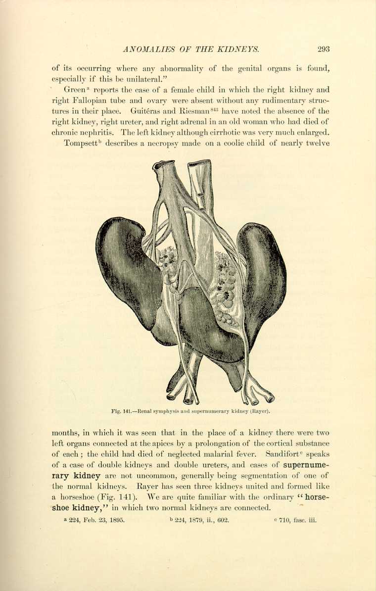
months, in which it was seen that in the place of a kidney there were two left organs connected at the apices by a prolongation of the cortical substance of each; the child had died of neglected malarial fever. Sandifort [6.318] speaks of a case of double kidneys and double ureters, and cases of supernumerary kidney are not uncommon, generally being segmentation of one of the normal kidneys. Rayer has seen three kidneys united and formed like a horseshoe (Fig. 141). We are quite familiar with the ordinary "horseshoe kidney,'' in which two normal kidneys are connected.
The kidney may be displaced to the pelvis, and Guinard *[390] quotes an instance in which the left kidney was situated in the pelvis, to the left of the rectum and back of the bladder. The ureter of the left side was very short. The left renal artery came from the bifurcation of the aorta and the primitive iliacs. The right kidney was situated normally, and received from the aorta two arteries, whose volume did not surpass the two arteries supplying the left suprarenal capsule, which was in its ordinary place. Displacements of the kidney anteriorly are very rare.
The ureters have been found multiple; Griffon [6.319] reports the history of a male subject in whom the ureter on the left side was double throughout its whole length; there were two vesical orifices on the left side one above the other; and Morestin, in the same journal, mentions ureters double on both sides in a female subject. Molinetti *[572] speaks of six ureters in one person. Littre in 1705 described a case of coalition of the ureters. Allen [6.320] describes an elongated kidney with two ureters. Coëyne [6.321] mentions duplication of the ureters on both sides. Lediberder [6.322] reports a case in which the ureter had double origin. Tyson [6.323] cites an instance of four ureters in an infant. Penrose [6.324] mentions the absence of the upper two-thirds of the left ureter, with a small cystic kidney, and there are parallel cases on record.
The ureters sometimes have anomalous terminations either in the rectum, vagina, or directly in the urethra. This latter disposition is realized normally in a number of animals and causes the incessant flow of urine, resulting in a serious inconvenience. Flajani speaks of the termination of the ureters in the pelvis; Nebel [6.325] has seen them appear just beneath the umbilicus; and Lieutaud describes a man who died at thirty-five, from another cause, whose ureters, as large as intestines, terminated in the urethral canal, causing him to urinate frequently; the bladder was absent. In the early part of this century [6.326] there was a young girl examined in New York whose ureters emptied into a reddish carnosity on the mons veneris. The urine dribbled continuously, and if the child cried or made any exertion it came in jets. The genital organs participated but little in the deformity, and with the exception that the umbilicus was low and the anus more anterior than natural, the child was well formed and its health good. Colzi [6.327] reports a case in which the left ureter opened externally at the left side of the hymen a little below the normal meatus urinarius. There is a case described [6.328] of a man who evidently suffered from a patent urachus, as the urine passed in jets
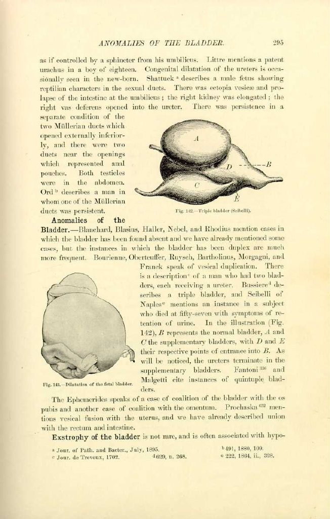
Fig. 142.—Triple bladder (Scibelli).
[Description: Drawing of a triple bladder]
Anomalies of the Bladder.—Blanchard, Blasius, Haller, Nebel, and Rhodius mention cases in which the bladder has been found absent and we have already mentioned some cases, but the instances in which the bladder has been duplex are much more frequent. Bourienne, Oberteuffer, Ruysch, Bartholinus, Morgagni, and Franck speak of vesical duplication. There is a description [6.331] of a man who had two bladders, each receiving a ureter. Bussiere [6.332] describes a triple bladder, and Scibelli of Naples [6.333] mentions an instance in a subject who died at fifty-seven with symptoms of retension of urine. In the illustration (Fig. 142), B represents the normal bladder, A and C the supplementary bladders, with D and E their respective points of entrance into B. As will be noticed, the ureters terminate in the supplementary bladders. Fantoni *[336] and

Fig. 143.—Dilation of the fetal bladder.
[Description: Drawing of dilation of the fetal bladder]
Malgetti cite instances of quintuple bladders.
The Ephemerides speaks of a case of coalition of the bladder with the os pubis and another case of coalition with the omentum. Prochaska *[652] mentions vesical fusion with the uterus, and we have already described union with the rectum and intestine.
Exstrophy of the bladder is not rare, and is often associated with hypospadias,
In an occasional instance in which there is occlusion at the umbilicus and again at the neck of the bladder this organ becomes so distended as to produce a most curious deformity in the fetus. Figure 143 shows such a case.
The Heart.—Absence of the heart has never been recorded in human beings except in the case of monsters, as, for example, the omphalosites, although there was a case reported and firmly believed by the ancient authors, —a Roman soldier in whom Telasius said he could discover no vestige of a heart. [6.334]
The absence of one ventricle has been recorded. Schenck [6.335] has seen the left ventricle deficient, and the Ephemerides, Behr, and Kerckring [6.336] speak of a single ventricle only in the heart. Riolan *[685] mentions a heart in which both ventricles were absent. Jurgens reported in Berlin, February 1, 1882, an autopsy on a child who had lived some days after birth, in which the left ventricle of the heart was found completely absent. Playfair [6.337] showed the heart of a child which had lived nine months in which one ventricle was absent. In King's College Hospital in London there is a heart of a boy of thirteen in which the cavities consist of a single ventricle and a single auricle.
Duplication of the heart, notwithstanding the number of cases reported, has been admitted with the greatest reserve by Geoffroy-Saint-Hilaire and by a number of authors. Among the celebrated anatomists who describe duplex heart are Littre, Meckel, Collomb, Panum, Behr, Paullini, Rhodins, Winslow, and Zacutus Lusitanus.
The Ephemerides [6.338] cites an instance of triple heart, and Johnston [6.339] has seen a triple heart in a goose.
The phenomenon of "blue-disease,'' or congenital cyanosis, is due to the patency of the foremen ovale, which, instead of closing at birth, persists sometimes to adult life.
Perhaps the most unique collection of congenital malformations of the heart from persons who have reached the age of puberty was to be seen in London in 1895. *[619] In this collection there was an adult heart in which the foremen ovale remained open until the age of thirty-seven; there were but two pulmonary valves; there was another heart showing a large patent foramen ovale from a man of forty-six; and there was a septum ventriculorum of an adult heart from a woman of sixty-three, who died of carcinoma of the breast, in which the foremen ovale was still open and would admit the forefinger.
Displacements of the heart are quite numerous. Deschamps of Laval made an autopsy on an old soldier which justified the expression, "He had a heart in his belly.'' This organ was found in the left lumbar region; it had, with its vessels, traversed an anomalous opening in the diaphragm. Franck observed in the Hospital of Colmar a woman with the heart in the epigastric region. Ramel [6.340] and Vetter speak of the heart under the diaphragm.
Inversion of the heart is quite frequent, and we often find reports of cases of this anomaly. Fournier [6.341] describes a soldier of thirty years, of middle height, well proportioned and healthy, who was killed in a duel by receiving a wound in the abdomen; postmortem, the heart was found in the position of the right lung; the two lungs were joined and occupied the left chest.
The anomalies of the vascular system are so numerous that we shall dismiss them with a slight mention. Malacarne in Torino in 1784 described a double aorta, and Hommelius [6.342] mentions an analogous case. The following case is quite an interesting anatomic anomaly: A woman since infancy had difficulty in swallowing, which was augmented at the epoch of menstruation and after exercise; bleeding relieved her momentarily, but the difficulty always returned. At last deglutition became impossible and the patient died of malnutrition. A necropsy revealed the presence of the subclavicular artery passing between the tracheal artery and the esophagus, compressing this latter tube and opposing the passage of food.
Anomalies of the Breasts.—The first of the anomalies of the generative apparatus to be discussed, although not distinctly belonging under this head, will be those of the mammæ.
Amazia, or complete absence of the breast, is seldom seen. Pilcher [6.343] describes an individual who passed for a female, but who was really a male, in whom the breasts were absolutely wanting. Foerster, Froriep, and Ried cite instances associated with thoracic malformation. Greenhow [6.344] reports a case in which the mammæ were absent, although there were depressed rudimentary nipples and areolæ. There were no ovaries and the uterus was congenitally imperfect.
There was a negress spoken of in 1842 in whom the right breast was missing, and there are cases of but one breast, mentioned by King, [6.345] Paull, [6.346] and others. [6.347] Scanzoni has observed absence of the left mamma with absence of the left ovary.
Polymazia is of much more frequent occurrence than is supposed. Julia, the mother of Alexander Severus, was surnamed "Mammea'' because she had supernumerary breasts. Anne Boleyn, the unfortunate wife of Henry VIII. of England, was reputed to have had six toes, six fingers, and three breasts. Lynceus says that in his time there existed a Roman woman with four mammæ, very beautiful in contour, arranged in two lines, regularly, one above the other, and all giving milk in abundance. Rubens has pictured a woman with four breasts; the painting may be seen in the Louvre in Paris.
There was a young and wealthy heiress who addressed herself to the ancient faculty at Tübingen, asking, as she displayed four mammary, whether, should she marry, she would have three or four children at a birth. This was a belief with which some of her elder matron friends had inspired her, and which she held as a hindrance to marriage.
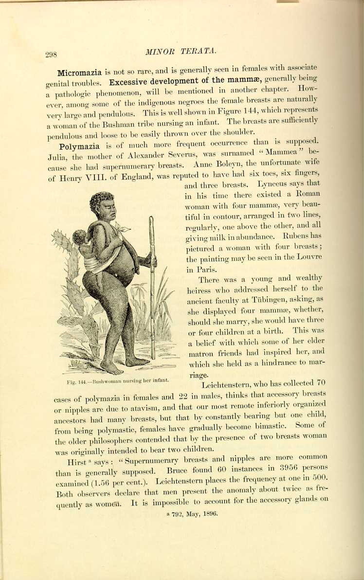
Fig. 144.—Bushwoman nursing her infant.
[Description: Drawing of a Bushwoman nursing her infant]
Leichtenstern, who has collected 70 cases of polymazia in females and 22 in males, thinks that accessory breasts or nipples are due to atavism, and that our most remote inferiorly organized ancestors had many breasts, but that by constantly bearing but one child, from being polymastic, females have gradually become bimastic. Some of the older philosophers contended that by the presence of two breasts woman was originally intended to bear two children.
Hirst [6.348] says: "Supernumerary breasts and nipples are more common than is generally supposed. Bruce found 60 instances in 3956 persons examined (1.56 per cent.). Leichtenstern places the frequency at one in 500. Both observers declare that men present the anomaly about twice as frequently as women. It is impossible to account for the accessory glands on
Possibly the greatest number of accessory mammæ reported is that of Neugebauer in 1886, who found ten in one person. Peuch in 1876 collected 77 cases, and since then Hamy, Quinqusud, Whiteford, Engstrom, and Mitchell Bruce have collected cases. Polymazia must have been known in the olden times, and we still have before us the old images of Diana, in which this goddess is portrayed with numerous breasts, indicating her ability to look after the growing child. Figure 145 shows an ancient Oriental statue of Artemisia or Diana now at Naples.
Bartholinus [6.349] has observed a Danish woman with three mammæ, two ordinarily formed and a third forming a triangle with the others and resembling the breasts of a fat man. In the village of Phullendorf in Germany early in this century there was an old woman who sought alms from place to place, exhibiting to the curious four symmetrical breasts, arranged parallel. She was extremely ugly, and when on all
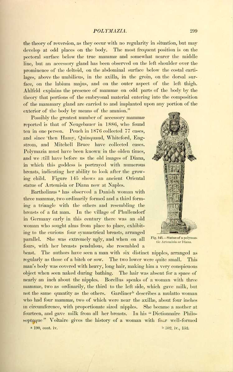
Fig. 145.—Statue of a Polymastic
Artemisisa or Diana.
[Description: Drawing of statue]
fours, with her breasts pendulous, she resembled a beast. The authors have seen a man with six distinct nipples, arranged as regularly as those of a bitch or sow. The two lower were quite small. This man's body was covered with heavy, long hair, making him a very conspicuous object when seen naked during bathing. The hair was absent for a space of nearly an inch about the nipples. Borellus speaks of a woman with three mammæ, two as ordinarily, the third to the left side, which gave milk, but not the same quantity as the others. Gardiner [6.350] describes a mulatto woman who had four mammæ, two of which were near the axillæ, about four inches in circumference, with proportionate sized nipples. She became a mother at fourteen, and gave milk from all her breasts. In his "Dictionnaire Philosophique'' Voltaire gives the history of a woman with four well-formed
Percy [6.351] says that among some prisoners taken in Austria was found a woman of Valachia, near Roumania, exceedingly fatigued, and suffering intensely from the cold. It was January, and the ground was covered with three feet of snow. She had been exposed with her two infants, who had been born twenty days, to this freezing temperature, and died on the next day. An examination of her body revealed five mammæ, of which four projected as ordinarily, while the fifth was about the size of that of a girl at puberty.
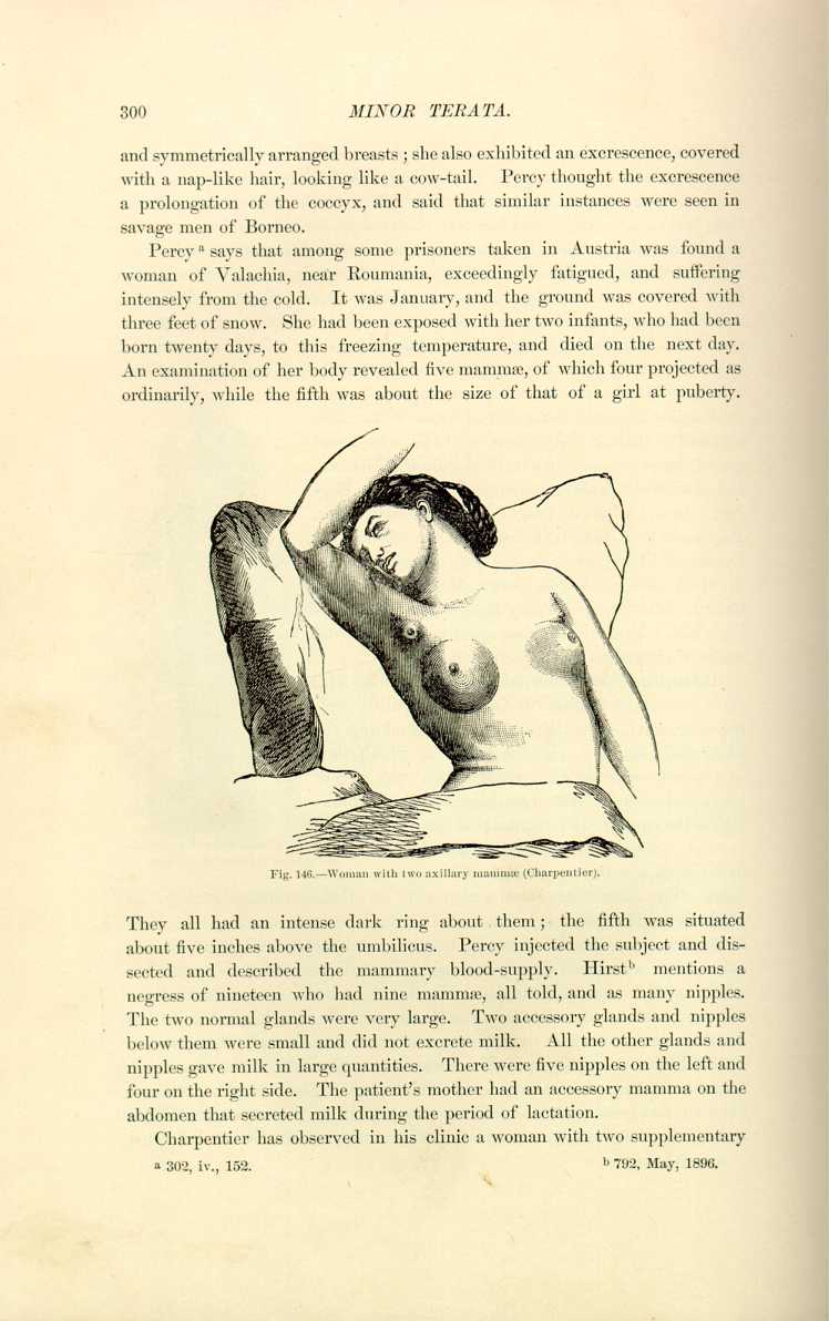
They all had an intense dark ring about them; the fifth was situated about five inches above the umbilicus. Percy injected the subject and dissected and described the mammary blood-supply. Hirst [6.352] mentions a negress of nineteen who had nine mammæ, all told, and as many nipples. The two normal glands were very large. Two accessory glands and nipples below them were small and did not excrete milk. All the other glands and nipples gave milk in large quantities. There were five nipples on the left and four on the right side. The patient's mother had an accessory mamma on the abdomen that secreted milk during the period of lactation.
Charpentier has observed in his clinic a woman with two supplementary
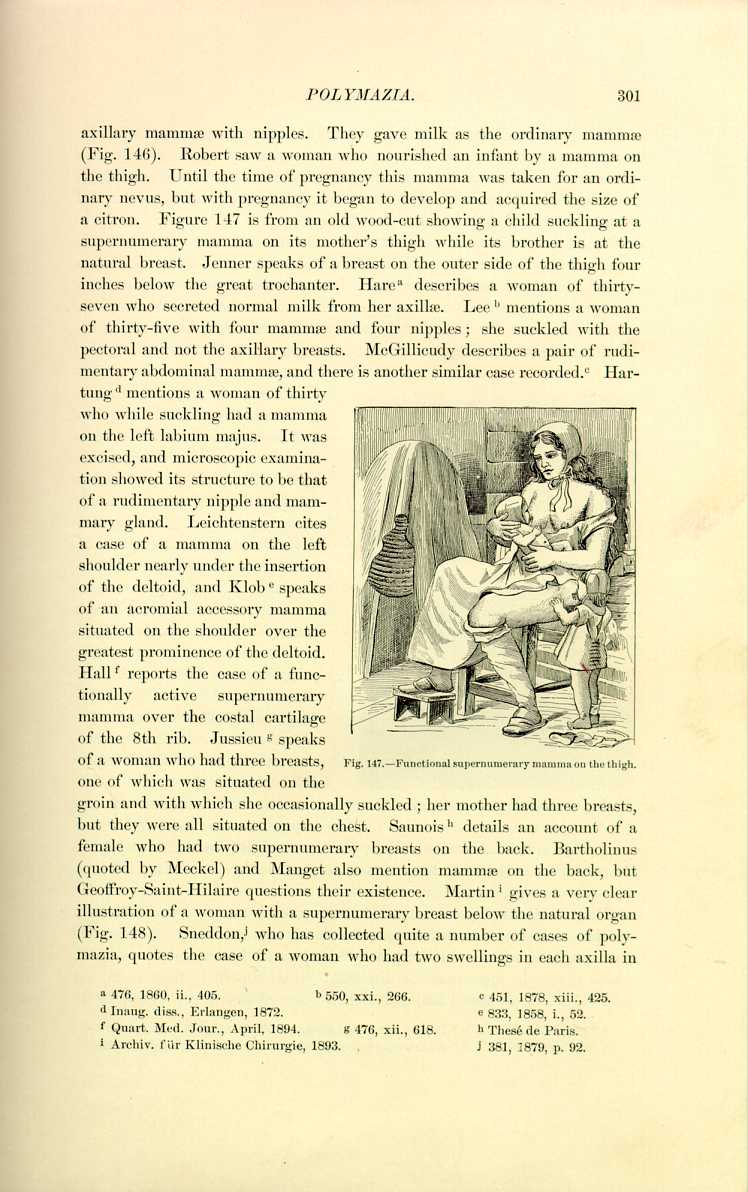
one of which was situated on the groin and with which she occasionally suckled; her mother had three breasts, but they were all situated on the chest. Saunois [6.360] details an account of a female who had two supernumerary breasts on the back. Bartholinus (quoted by Meckel) and Manget also mention mammæ on the back, but Geoffroy-Saint-Hilaire questions their existence. Martin [6.361] gives a very clear illustration of a woman with a supernumerary breast below the natural organ (Fig. 148). Sneddon, [6.362] who has collected quite a number of cases of polymazia, quotes the case of a woman who had two swellings in each axilla in
Polymazia sometimes seems to be hereditary. Robert saw a daughter whose mother was polymastic, and Woodman [6.363] saw a mother and eldest daughter who each had three nipples. Lousier [6.364] mentions a woman wanting a mamma who transmitted this vice of conformation to her daughter. Handyside says he knew two brothers in both of whom breasts were wanting.
Supernumerary nipples alone are also seen, as many as five having been found on the same breast. Neugebauer reports eight supernumerary nipples in one case. Hollerus has seen a woman who had two nipples on the same breast which gave milk with the same regularity and the same abundance as the single nipple. The Ephemerides contains a description of a triple nipple. Barth [6.365] describes "mamma erratica'' on the face in front of the right ear which enlarged during menstruation.
Cases of deficiency of the nipples have been reported by the Ephemerides, Lentilius, Severinus, and Werckardus.
Cases of functional male mammæ will be discussed in Chapter IX.
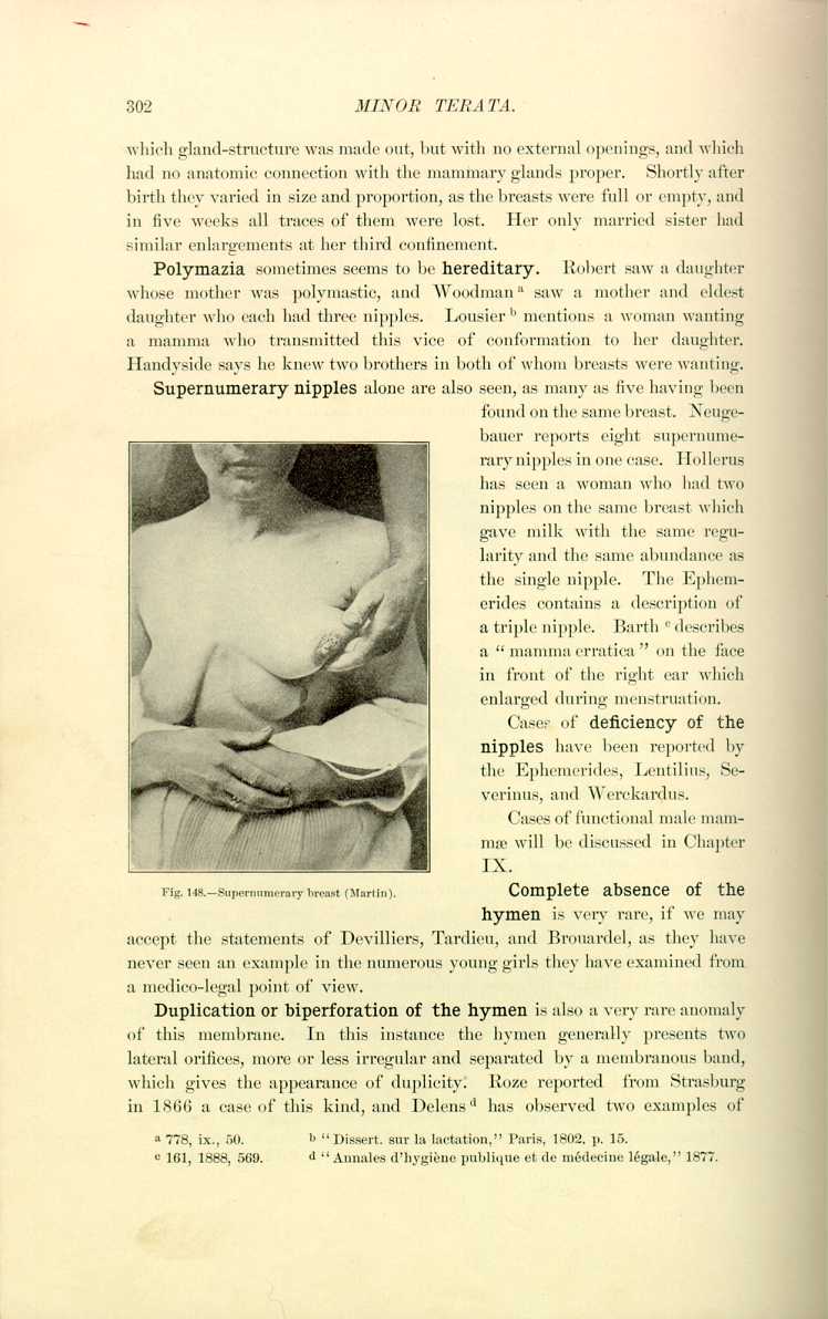
Fig. 143.—Supernumerary breast (Martin).
[Description: Photograph of a supernumerary breast]
Complete absence of the hymen is very rare, if we may accept the statements of Devilliers, Tardieu, and Brouardel, as they have never seen an example in the numerous young girls they have examined from a medico-legal point of view.
Duplication or biperforation of the hymen is also a very rare anomaly of this membrane. In this instance the hymen generally presents two lateral orifices, more or less irregular and separated by a membranous band, which gives the appearance of duplicity. Roze reported from Strasburg in 1866 a case of this kind, and Delens [6.366] has observed two examples of
The different varieties of the hymen will be left to the works on obstetrics. As has already been observed, labor is frequently seriously complicated by a persistent and tough hymen.
Deficient vulva may be caused by the persistence of a thick hymen, by congenital occlusion, or by absolute absence in vulvar structure. Bartholinus, Borellus, Ephemerides, Julius, Vallisneri, and Baux are among the older writers who mention this anomaly, but as it is generally associated with congenital occlusion, or complete absence of the vagina, the two will be considered together.
Complete absence of the vagina is quite rare. Baux [6.367] reports a case of a girl of fourteen in whom "there was no trace of fundament or of genital organs.'' Oberteuffer [6.368] speaks of a case of absent vagina. Vicq d'Azir [6.369] is accredited with having seen two females who, not having a vagina, copulated all through life by the urethra, and Fournier sagely remarks that the extra large urethra may have been a special dispensation of nature. Bosquet [6.370] describes a young girl of twenty with a triple vice of conformation—an obliterated vulva, closure of the vagina, and absence of the uterus. Menstrual hemorrhage took place from the gums. Clarke [6.371] has studied a similar case which was authenticated by an autopsy.
O'Ferral of Dublin, Gooch, Davies, Boyd, Tyler Smith, Hancock, Coste, Klayskens, Debrou, Braid, Watson, and others are quoted by Churchill as having mentioned the absence of the vagina. Amussat [6.372] observed a German girl who did not have a trace of a vagina and who menstruated regularly. Griffith [6.373] describes a specimen in the Museum of St. Bartholomew's Hospital, London, in which the ovaries lay on the surface of the pelvic peritoneum and there was neither uterus nor vagina; the pelvis had some of the characteristics of the male type. Matthews Duncan has observed a somewhat similar case, the vagina not measuring more than an inch in length. Ferguson [6.374] describes a prostitute of eighteen who had never menstruated. The
Brodhurst [6.375] reports an instance of absence of the vagina and uterus in a girl of sixteen who at four years of age showed signs of approaching puberty. At this early age the mons was covered with hair, and at ten the clitoris was three inches long and two inches in circumference. The mammæ were well developed. The labia descended laterally and expanded into folds, resembling the scrotum.
Azéma [6.376] reports an instance of complete absence of the vagina and impermeability and probable absence of the col uterinus. The deficiencies were remedied by operation. Bérard [6.377] mentions a similar deformity and operation in a girl of eighteen. Gooding [6.378] cites an instance of absent vagina in a married woman, the uterus discharging the functions. Gosselin [6.379] reports a case in which a voluminous tumor was formed by the retained menstrual fluid in a woman without a vagina. An artificial vagina was created, but the patient died from extravasation of blood into the peritoneal cavity. Carter, Polaillon, Martin, Curtis, Worthington, Hall, Hicks, Molière, Patry, Dolbeau, Desormeaux, and Gratigny also record instances of absence of the vagina.
There are some cases reported in extramedical literature which might be cited. Bussy Rabutin in his Mémoires in 1639 speaks of an instance. The celebrated Madame Récamier was called by the younger Dumas an involuntary virgin; and in this connection could be cited the malicious and piquant sonnet—
CHATEAUBRIAND ET MADAME RéCAMIER.
"Juliette et René s'aimaient d'amour si tendreQue Dien, sans les punir, a pu leur pardonner:
Il n'avait pas voulu que l'une pût donner
Ce que l'autre ne pouvait prendre.''
Duplex vagina has been observed by Bartholinus, Malacarne, Asch, Meckel, Osiander, Purcell, and other older writers. In more modern times reports of this anomaly are quite frequent. Hunter [6.380] reports a case of labor at the seventh month in a woman with a double vagina, and delivery through the rectum. Atthill and Watts speak of double vagina with single uterus.
Cullingworth [6.384] cites two cases in which there were transverse septa of the vagina. Stone [6.385] reports five cases of transverse septa of the vagina. Three of the patients were young women who had never borne children or suffered injury. Pregnancy existed in each case. In the first the septum was about two inches from the introitus, and contained an opening about 1/2 inch in diameter which admitted the tip of the finger. The membrane was elastic and thin and showed no signs of inflammation. Menstruation had always been regular up to the time of pregnancy. The second was a duplicate of the first, excepting that a few bands extended from the cervix to the membranous septum. In the third the lumen of the vagina, about two inches from the introitus, was distinctly narrowed by a ridge of tissue. There was uterine displacement and some endocervicitis, but no history of injury or operation and no tendency to contraction. The two remaining cases occurred in patients seen by Dr. J. F. Scott. In one the septum was about 1 3/4 inches from the entrance to the vagina and contained an orifice large enough to admit a uterine probe. During labor the septum resisted the advance of the head for several hours, until it was slit in several directions. In the other, menstruation had always been irregular, intermissions being followed by a profuse flow of black and tarry blood, which lasted sometimes for fifteen days and was accompanied by severe pain. The septum was 1 1/2 inches from the vaginal orifice and contained an opening which admitted a uterine sound. It was very dense and tight and fully 1/8 inch in thickness.
Mordie [6.386] reported a case of congenital deficiency of the rectovaginal septum which was successfully remedied by operation.
Anomalous Openings of the Vagina.—The vagina occasionally opens abnormally into the rectum, into the bladder, the urethra, or upon the
Opening of the vagina upon the parietes, of which Le Fort has collected a number of cases, has never been observed in connection with a viable fetus.
Absence of the labia majora has been observed, especially by Pozzi, to the exclusion of all other anomalies. It is the rule in exstrophy of the bladder.
Absence of the nymphæ has also been observed, particularly by Auvard and by Perchaux, and is generally associated with imperfect development of the clitoris. Constantinedes [6.389] reports absence of the external organs of generation, probably also of the uterus and its appendages, in a young lady. Van Haartman, LeFort, Magee, and Ogle cite cases of absence of the external female organs. Riolan [6.390] in the early part of the seventeenth century reported a case of defective nymphæ; Neubauer in 1774 offers a contrast to this case in an instance of triple nymphæ.
The nymphæ are sometimes enormously enlarged by hypertrophy, by varicocele, or by elephantiasis, of which latter type Rigal de Gaillac has observed a most curious case. There is also a variety of enlargement of the clitoris which seems to be constant in some races; it may be a natural hypertrophy, or perhaps produced by artificial manipulation.
The peculiar conditions under which the Chinese women are obliged to live, particularly their mode of sitting, is said to have the effect of causing unusual development of the mons veneris and the labia majora. On the other hand, some of the lower African races have been distinguished by
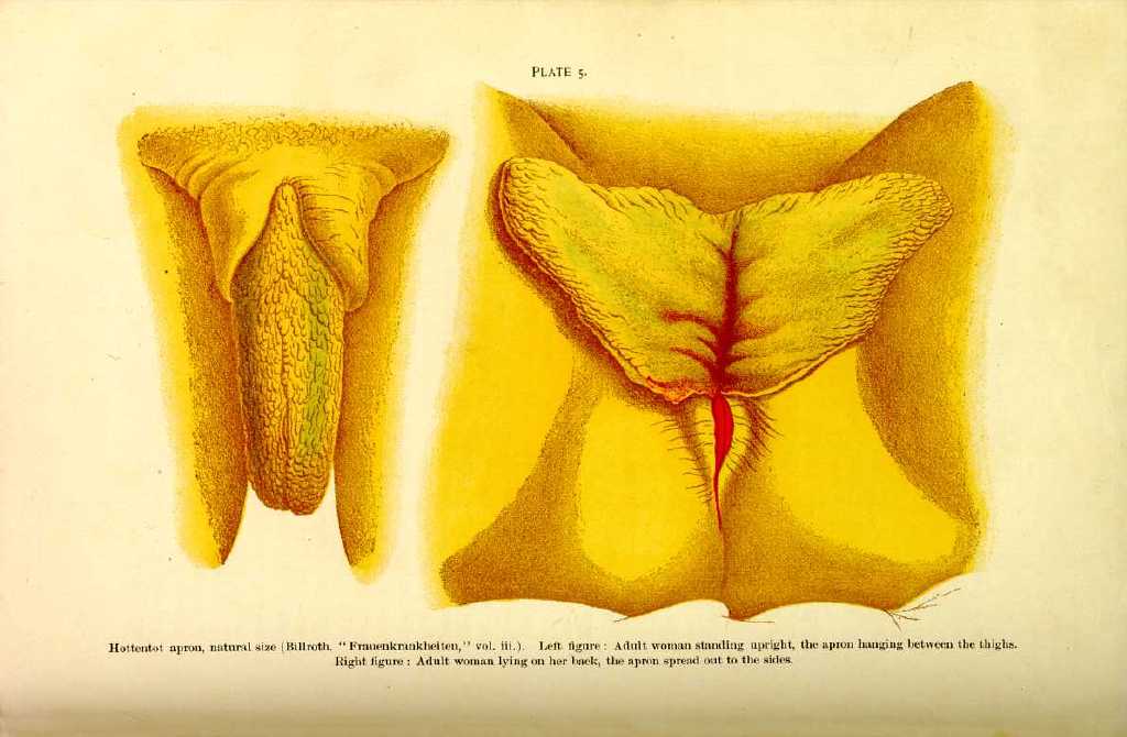
The labial appendages of the Hottentot female have been celebrated for many years. Blumenbach and others of the earlier travelers found that the apron-like appearance of the genitals of the Hottentot women was due to abnormal hypertrophy of the labia and nymphæ (Pl. 5). According to John Knott, the French traveler, Le Vaillant, said that the more coquettish among the Hottentot girls are excited by extreme vanity to practice artificial elongation of the nympha and labia. They are said to pull and rub these parts, and even to stretch them by hanging weights to them. Some of them are said to spend several hours a day at this process, which is considered one of the important parts of the toilet of the Hottentot belle, this malformation being an attraction for the male members of the race. Merensky says that in Basutoland the elder women begin to practice labial manipulation on their female children shortly after infancy, and Adams has found this custom to prevail in Dahomey; he says that the King's seraglio includes 3000 members, the elect of his female subjects, all of whom have labia up to the standard of recognized length. Cameron found an analogous practice among the women of the shores of Lake Tanganyika. The females of this nation manipulated the skin of the lower part of the abdomens of the female children from infancy, and at puberty these women exhibit a cutaneous curtain over the genitals which reaches half-way down the thighs.
A corresponding development of the preputian clitorides, attaining the length of 18 mm. or even more, has been observed among the females of Bechuanaland. The greatest elongation measured by Barrow was five inches, but it is quite probable that it was not possible for him to examine the longest, as the females so gifted generally occupied very high social positions.
Morgagni describes a supernumerary left nympha, and Petit is accredited with seeing a case which exhibited neither nymphæ, clitoris, nor urinary meatus. Mauriceau performed nymphotomy on a woman whose nymphæ were so long as to render coitus difficult. Morand *[575] quotes a case of congenital malformation of the nymphæ, to which he attributed impotency.
There is sometimes coalition of the labia and nymphæ, which may be so firm and extensive as to obliterate the vulva. Debout [6.391] has reported a case of absence of the vulva in a woman of twenty upon whom he operated, which was the result of the fusion of the labia minora, and this with an enlarged clitoris gave the external appearance of an hermaphrodite.
The absence of the clitoris coincides with epispadias in the male, and in atrophy of the vulva it is common to find the clitoris rudimentary; but a more frequent anomaly is hypertrophy of the clitoris.
We learn from the classics that there were certain females inhabiting the borders of the ægean Sea who had a sentimental attachment for one another which was called "Lesbian love,'' and which carried them to the highest degree of frenzy. The immortal effusions of Sappho contain references to this passion. The solution of this peculiar ardor is found in the fact that some of the females had enlarged clitorides, strong voices, robust figures, and imitated men. Their manner was imperative and authoritative to their sex, who worshiped them with perverted devotion. We find in Martial [6.393] mention of this perverted love, and in the time of the dissolute Greeks and Romans ridiculous jealousies for unfaithfulness between these women prevailed. Aetius said that the Egyptians practiced amputation of the clitoris, so that enlargement of this organ must have been a common vice of conformation along the Nile. It was also said that the Egyptian women practiced circumcision on their females at the age of seven or eight, the time chosen being when the Nile was in flood. Bertherand [6.394] cites examples of enlarged clitorides in Arab women; Bruce testifies to this circumstance in Abyssinia, and Mungo Park has observed it in the Mandingos and the Ibbos.
Sonnini [6.395] says that the women of Egypt had a natural excrescence, fleshy in consistency, quite thick and pendulous, coming from the skin of the mons veneris. Sonnini says that in a girl of eight he saw one of these caruncles which was 1/2 inch long, and another on a woman of twenty which was four inches long, and remarks that they seem peculiar only to women of distinct Egyptian origin.
Duhouset [6.396] says that in circumcision the Egyptian women not only remove a great part of the body of the clitoris with the prepuce, but also adjacent portions of the nymphæ; Gallieni [6.397] found a similar operation customary on the upper banks of the Niger.
Otto at Breslau in 1824 reports seeing a negress with a clitoris 4 1/2 inches long and 1 1/2 inches in the transverse diameter; it projected from the vulva and when supine formed a complete covering for the vaginal orifice. The clitoris may at times become so large as to prevent coitus, and in France has constituted a legitimate cause for divorce. This organ is very sensitive, and it is said that in cases of supposed catalepsy a woman cannot bear titillation of the clitoris without some visible movement.
Columbus cites an example of a clitoris as long as a little finger; Haller
In some cases ossification of the clitoris is observed Fournier *[302] speaks of a public woman in Venice who had an osseous clitoris; it was said that men having connection with her invariably suffered great pain, followed by inflammation of the penis.
There are a few instances recorded of bifid clitoris,
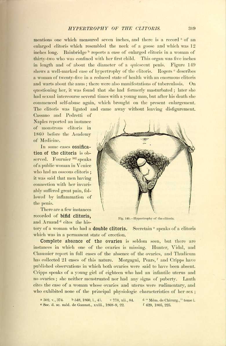
Fig. 149.—Hypertrophy of the clitoris.
[Description: Drawing showing hypertrophy of the clitoris]
and Arnaud [6.401] cites the history of a woman who had a double clitoris. Secretain [6.402] speaks of a clitoris which was in a permanent state of erection.
Complete absence of the ovaries is seldom seen, but there are instances in which one of the ovaries is missing. Hunter, Vidal, and Chaussier report in full cases of the absence of the ovaries, and Thudicum has collected 21 cases of this nature. Morgagni, Pears, [6.403] and Cripps have published observations in which both ovaries were said to have been absent. Cripps speaks of a young girl of eighteen who had an infantile uterus and no ovaries; she neither menstruated nor had any signs of puberty. Lauth cites the case of a woman whose ovaries and uterus were rudimentary, and who exhibited none of the principal physiologic characteristics of her sex;
At one time the existence of genuine supernumerary ovaries was vigorously disputed, and the older records contain no instances, but since the researches of Beigel, Puech, Thudicum, Winckler, de Sinéty, and Paladino the presence of multiple ovaries is an incontestable fact. It was originally thought that supernumerary ovaries as well as supernumerary kidneys were simply segmentations of the normal organs and connected to them by portions of the proper substance; now, however, by the recent reports we are warranted in admitting these anomalous structures as distinct organs. It has even been suggested that it is the persistence of these ovaries that causes the menstruation of which we sometimes hear as taking place after ovariotomy. Sippel [6.404] records an instance of third ovary; Mangiagalli [6.405] has found a supernumerary ovary in the body of a still-born child, situated to the inner side of the normal organ. Winckel discovered a large supernumerary ovary connected to the uterus by its own ovarian ligament. Klebs found two ovaries on one side, both consisting of true ovarian tissue, and connected by a band 3/5 inch long.
Doran divides supernumerary ovaries into three classes:—
(1) The ovarium succentauriatum of Beigel.
(2) Those cases in which two masses of ovarian tissue are separated by ligamentous bands.
(3) Entirely separate organs, as in Winckel's case.
Prolapsus or displacement of the ovaries into the culdesac of Douglas, the vaginal wall, or into the rectum can be readily ascertained by the resulting sense of nausea, particularly in defecation or in coitus. Mundé, Barnes, Lentz, Madden, and Heywood Smith report instances, and Cloquet describes an instance of inguinal hernia of the ovary in which the uterus as well as the Fallopian tube were found in the inguinal canal. Debierre [6.406] mentions that Puech has gathered 88 instances of inguinal hernia of the ovary and 14 of the crural type, and also adds that Otte [6.407] cites the only instance in which crural ovarian hernia has been found on both sides. Such a condition with other associate malformations of the genitalia might easily be mistaken for an instance of hermaphroditic testicles.
The Fallopian tubes are rarely absent on either side, although Blasius *[214] reports an instance of deficient oviducts. Blot [6.408] reports a case of atrophy, or rather rudimentary state of one of the ovaries, with absence of the tube on that side, in a woman of forty.
Doran [6.409] has an instance of multiple Fallopian tubes, and Richard, in 1861, says several varieties are noticed. These tubes are often found fused
Absence of the uterus is frequently reported. Lieutaud and Richerand *[302] are each said to have dissected female subjects in whom neither the uterus nor its annexed organs were found. Many authors are accredited with mentioning instances of defective or deficient uteri, among them Bosquet, [6.410] Boyer, [6.411] Walther, *[814] Le Fort, Calori, Pozzi, Mundé, and Strauch. Balade [6.412] has reported a curious absence of the uterus and vagina in a girl of eighteen. Azem, Bastien, Bibb, Bovel, Warren, Ward, and many others report similar instances, and in several cases all the adnexa as well as the uterus and vagina were absent, and even the kidney and bladder malformed.
Phillips [6.413] speaks of two sisters, both married, with congenital absence of the uterus. In his masterly article on "Heredity,'' [6.414] Sedgwick quotes an instance of total absence of the uterus in three out of five daughters of the same family; two of the three were twice married.
Double uterus is so frequently reported that an enumeration of the cases would occupy several pages. Bicorn, bipartite, duplex, and double uteruses are so called according to the extent of the duplication. The varieties range all the way from slight increase to two distinct uteruses, with separate appendages and two vaginæ. Meckel, Boehmer, and Callisen are among the older writers who have observed double uterus with associate double vagina. Figure 150 represents a transverse section of a bipartite uterus with a double vagina. The so-called uterus didelphus is really a duplex uterus, or a veritable double uterus, each segment having the appearance of a complete unicorn uterus more or less joined to its neighbor (Fig. 151). Vallisneri [6.415] relates the history of a woman who was poisoned by cantharides who had two uteruses, one opening into the vagina, the other into the rectum. Morand, Bartholinus, Tiedemann, Ollivier, Blundell, and many others relate instances of double uterus in which impregnation had occurred, the fetus being retained until the full term.
Purcell of Dublin [6.416] says that in the summer of 1773 he opened the body of a woman who died in the ninth month of pregnancy. He found a uterus of ordinary size and form as is usual at this period of gestation, which contained a full-grown fetus, but only one ovary attached to a single Fallopian tube. On the left side he found a second uterus, unimpregnated and of usual size, to which another ovary and tube were attached. Both of these uteruses were distinct and almost entirely separate.
Pregnancy with Double Uterus.—Hollander [6.417] describes the following anomaly of the uterus which he encountered during the performance of a celiotomy:—
"There were found two uteruses, the posterior one being a normal organ

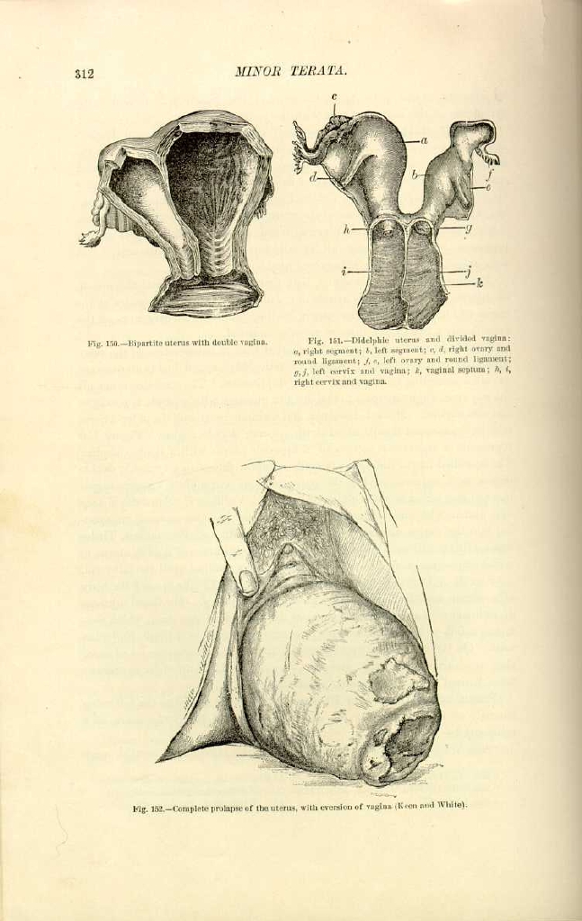

Other cases of double uterus with pregnancy are mentioned on page 49.
When there is simultaneous pregnancy in each portion of a double uterus a complication of circumstances arises. Debierre quotes an instance of a woman who bore one child on July 16, 1870, and another on October 31st of the same year, and both at full term. She had only had three menstrual periods between the confinements. The question as to whether a case like this would be one of superfetation in a normal uterus, or whether the uterus was double, would immediately arise. There would also be the possibility that one of the children was of protracted gestation or that the other was of premature birth. Article 312 of the Civil Code of France accords a minimum of one hundred and eighty and a maximum of three hundred days for the gestation of a viable child. (See Protracted Gestation.)
Voight [6.418] is accredited with having seen a triple uterus, and there are several older parallels on record. Thilow mentions a uterus which was
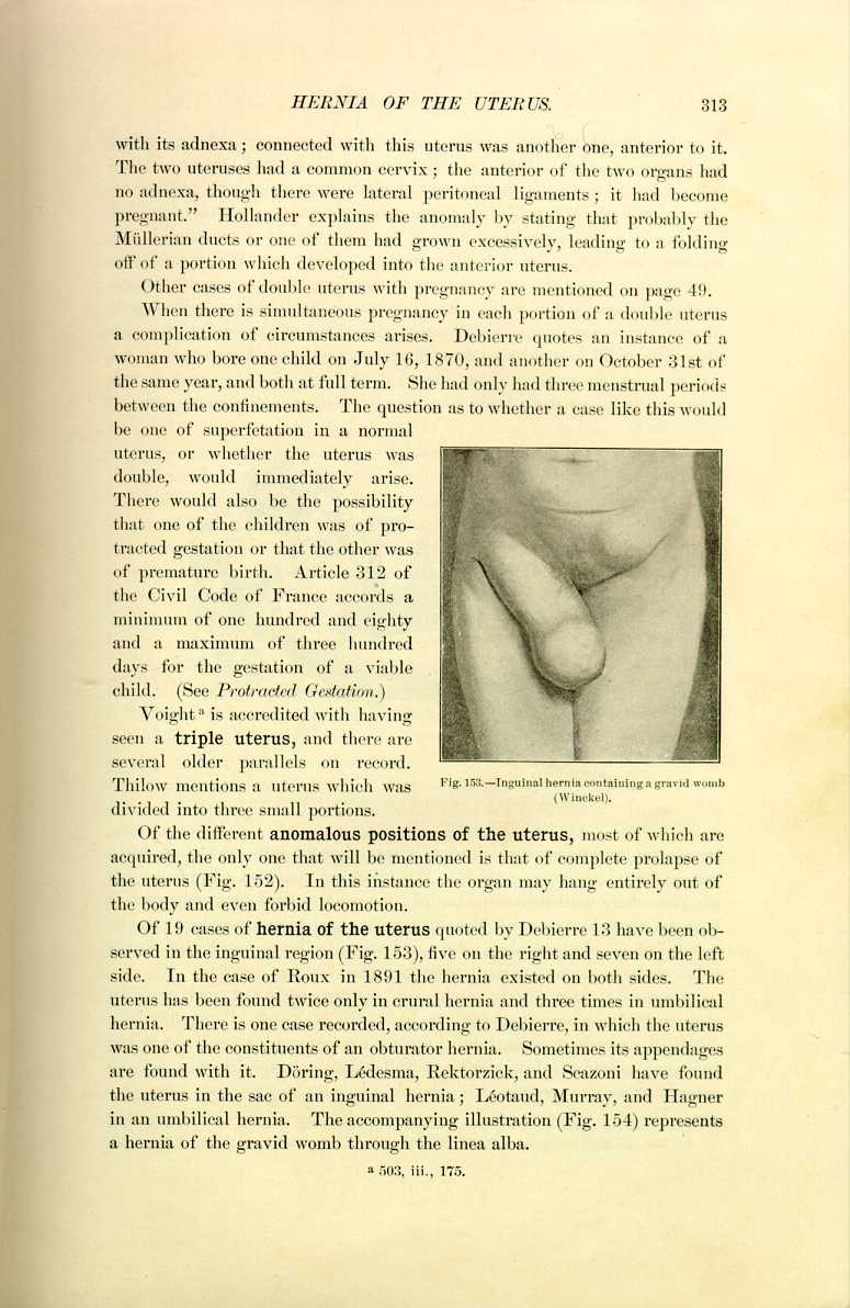
divided into three small portions.
Of the different anomalous positions of the uterus, most of which are acquired, the only one that will be mentioned is that of complete prolapse of the uterus (Fig. 152). In this instance the organ may hang entirely out of the body and even forbid locomotion.
Of 19 cases of hernia of the uterus quoted by Debierre 13 have been observed in the inguinal region (Fig. 153), five on the right and seven on the left side. In the case of Roux in 1891 the hernia existed on both sides. The uterus has been found twice only in crural hernia and three times in umbilical hernia. There is one case recorded, according to Debierre, in which the uterus was one of the constituents of an obturator hernia. Sometimes its appendages are found with it. Döring, Lédesma, Rektorzick, and Scazoni have found the uterus in the sac of an inguinal hernia; Léotaud, Murray, and Hagner in an umbilical hernia. The accompanying illustration (Fig. 154) represents a hernia of the gravid womb through the linea alba.
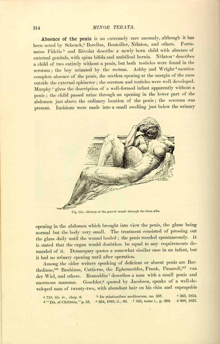
opening in the abdomen which brought into view the penis, the glans being normal but the body very small. The treatment consisted of pressing out the glans daily until the wound healed; the penis receded spontaneously. It is stated that the organ would doubtless be equal to any requirements demanded of it. Demarquay quotes a somewhat similar case in an infant, but it had no urinary opening until after operation.
Among the older writers speaking of deficient or absent penis are Bartholinus, *[190] Bauhinus, Cattierus, the Ephemerides, Frank, Panaroli, *[617] van der Wiel, and others. Renauldin [6.424] describes a man with a small penis and enormous mammæ. Goschler, [6.425] quoted by Jacobson, speaks of a well-developed man of twenty-two, with abundant hair on his chin and suprapubic
Many instances of rudimentary development of the penis have been recorded, most of them complicated with cryptorchism or other abnormality of the sexual organs. In other instances the organ is present, but the infantile type is present all through life; sometimes the subjects are weak in intellect and in a condition similar to cretinism. Kaufmann quotes a case in a weakly boy of twelve whose penis was but 3/4 inch long, about as thick as a goose-quill, and feeling as limp as a mere tube of skin; the corpora cavernosa were not entirely absent, but ran only from the ischium to the junction of the fixed portion of the penis, suddenly terminating at this point. Nothing indicative of a prostate could be found. The testicles were at the entrance of the inguinal canal and the glans was only slightly developed.
Binet [6.426] speaks of a man of fifty-three whose external genitalia were of the size of those of a boy of nine. The penis was of about the size of the little finger, and contained on each side testicles not larger than a pea. There was no hair on the pubes or the face, giving the man the aspect of an old woman. The prostate was almost exterminated and the seminal vesicles were very primitive in conformation. Wilson was consulted by a gentleman of twenty-six as to his ability to perform the marital function. In size his penis and testicles hardly exceeded those of a boy of eight. He had never felt desire for sexual intercourse until he became acquainted with his intended wife, since when he had erections and nocturnal emissions. The patient married and became the father of a family; those parts which at twenty-six were so much smaller than usual had increased at twenty-eight to normal adult size. There are three cases on record in the older literature of penises extremely primitive in development. They are quoted by the Ephemerides, Plater, *[635] Schenck, [6.427] and Zacchias. The result in these cases was impotency.
In the Army and Medical Museum at Washington are two injected specimens of the male organ divested of skin. From the meatus to the pubis they measure 6 1/2 and 5 1/2 inches; from the extremity to the termination of either crus 9 3/4 and 8 3/4 inches, and the circumferences are 4 3/4 and 4 1/4 inches. Between these two we can strike an average of the size of the normal penis.
The pénis palmé, or suture de la verge of the French, is the name given to those examples of single cutaneous envelope for both the testicles and penis; the penis is adherent to the scrotum by its inferior face; the glans only is free and erection is impossible. Chrétien cites an instance in a man of twenty-five, and Schrumpf of Wesserling [6.431] describes an example of this rare anomaly. The penis and testes were inclosed in a common sac, a slight projection not over 1/4 inch long being seen from the upper part of this curious scrotum. When the child was a year old a plastic operation was performed on this anomalous member with a very satisfactory result. Petit describes an instance in which the penis was slightly fused with the scrotum.
There are many varieties of torsion of the penis. The glans itself may be inclined laterally, the curvature may be total, or there may be a veritable rotation, bringing the inferior face above and the superior face below. Gay [6.432] describes a child with epispadias whose penis had undergone such torsion on its axis that its inferior surface looked upward to the left, and the child passed urine toward the left shoulder. Follin [6.433] mentions a similar instance in a boy of twelve with complete epispadias, and Verneuil and Guerlin also record cases, both complicated with associate maldevelopment. Caddy [6.434] mentions a youth of eighteen who had congenital torsion of the penis with out hypospadias or epispadias. There was a complete half-turn to the left, so that the slit-like urinary meatus was reversed and the frenum was above. Among the older writers who describe incurvation or torsion of the penis are Arantius, [6.435] the Ephemerides, Haenel, *[401] Petit, [6.436] Schurig, Tulpius, [6.437] and Zacchias. *[830]
Zacutus Lusitans [6.438] speaks of torsion of the penis from freezing. Paullini [6.439] mentions a case the result of masturbation, and Hunter [6.440] speaks of torsion of the penis associated with arthritis.
Ossification of the Penis.—MacClellann [6.441] speaks of a man of fifty-two whose penis was curved and distorted in such a manner that urine could not be passed without pain and coitus was impossible. A bony mass was discovered in the septum between the corpora cavernosa; this was dissected out with much hemorrhage and the upward curvature was removed, but there
The complete absence of the frenum and prepuce has been observed in animals but is very rare in man. The incomplete or irregular development is more frequent, but most common is excessive development of the prepuce, constituting phimosis, when there is abnormal adherence with the glans. Instances of phimosis, being quite common, will be passed without special mention. Deficient or absent prepuce has been observed by Blasius, *[214] Marcellus Donatus, *[306] and Gilibert. Partial deficiency is described by Petit Severinus, and others.
There may be imperforation or congenital occlusion of some portion of the urethra, causing enormous accumulation of urine in the bladder, but fortunately there is generally in such cases some anomalous opening of the urethra giving vent to the excretions. Tulpius [6.443] mentions a case of deficient urethra. In the Ephemerides there is an account of a man who had a constant flow of semen from an abnormal opening in the abdomen. La Peyroma [6.444] describes a case of impotence due to ejaculation of the spermatic ducts into the bladder instead of into the urethra, but remarks that there was a cicatrix of a wound of the neighboring parts. There are a number of instances in which the urethra has terminated in the rectum. Congenital dilatation of the urethral canal is very rare, and generally accompanied by other malformation.
Duplication of the urethra or the existence of two permeable canals is not accepted by all the authors, some of whom contend that one of the canals either terminates in a culdesac or is not separate in itself. Verneuil has published an article clearly exposing a number of cases, showing that it is possible for the urethra to have two or more canals which are distinct and have separate functions. Fabricius Hildanus [6.445] speaks of a double aperture to the urethra; Marcellus Donatus [6.446] describes duplicity of the urethra, one of the apertures being in the testicle; and there is another case on record [6.447] in which there was a urethral aperture in the groin. A case of double urethra in a man of twenty-five living in Styria [6.448] who was under treatment for gonorrhea is described, the supernumerary urethra opening above the natural one and receiving a sound to the depth of 17 cm. There was purulent gonorrhea in both urethræ. Vesalius [6.449] has an account of a double urethral aperture, one of which was supposed to give spermatic fluid and the other urine. Borellus, Testa, and Cruveilhier have reported similar instances. Instances of double penis have been discussed under the head of diphallic terata, page 194.
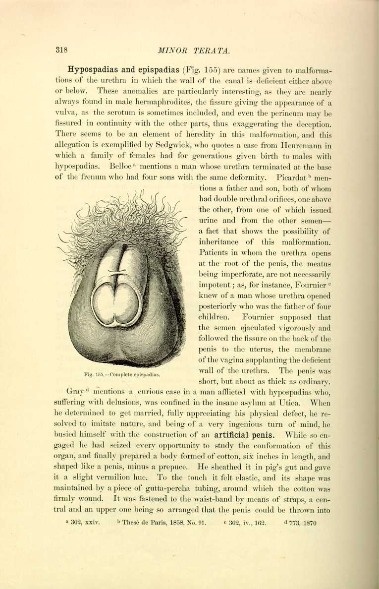
Fig. 155.—Complete epispadias.
[Description: Drawing of complete epispadias]
Gray [6.453] mentions a curious case in a man afflicted with hypospadias who, suffering with delusions, was confined in the insane asylum at Utica. When he determined to get married, fully appreciating his physical defect, he resolved to imitate nature, and being of a very ingenious turn of mind, he busied himself with the construction of an artificial penis. While so engaged he had seized every opportunity to study the conformation of this organ, and finally prepared a body formed of cotton, six inches in length, and shaped like a penis, minus a prepuce. He sheathed it in pig's gut and gave it a slight vermilion hue. To the touch it felt elastic, and its shape was maintained by a piece of gutta-percha tubing, around which the cotton was firmly wound. It was fastened to the waist-band by means of straps, a central and an upper one being so arranged that the penis could be thrown into
Entire absence of the male sexual apparatus is extremely rare, but Blondin and Velpeau have reported cases.
Complete absence of the testicles, or anorchism, is a comparatively rare anomaly, and it is very difficult to distinguish between anorchism and arrest of development, or simple atrophy, which is much more common. Fisher of Boston [6.457] describes the case of a man of forty-five, who died of pneumonia. From the age of puberty to twenty-five, and even to the day of death, his voice had never changed and his manners were decidedly effeminate. He always sang soprano in concert with females. After the age of twenty-five, however, his voice became more grave and he could not accompany females with such ease. He had no beard, had never shaved, and had never exhibited amorous propensities or desire for female society. When about twenty-one he became associated with a gay company of men and was addicted to the cup, but would never visit houses of ill-fame. On dissection no trace of testicles could be found; the scrotum was soft and flabby. The cerebellum was the exact size of that of a female child.
Individuals with one testicle are called monorchids, and may be divided into three varieties:—
(1) A solitary testicle divided in the middle by a deep fissure, the two lobes being each provided with a spermatic cord on the same side as the lobe.
(2) Testicles of the same origin, but with coalescence more general.
(3) A single testicle and two cords.
Gruber of St. Petersburg [6.458] held a postmortem on a man in January,
Polyorchids are persons who have more than two testicles. For a long time the abnormality was not believed to exist, and some of the observers denied the proof by postmortem examination of any of the cases so diagnosed, but there is at present no doubt of the fact,—three, four, and five testicles having been found at autopsies. Russell, one of the older writers on the testicle, mentions a monk who was a triorchid, and was so salacious that his indomitable passion prevented him from keeping his vows of chastity. The amorous propensities and generative faculties of polyorchids have always been supposed greater than ordinary. Russell reports another case of a man with a similar peculiarity, who was prescribed a concubine as a reasonable allowance to a man thus endowed.
Morgagni and Meckel say that they never discovered a third testicle in dissections of reputed triorchids, and though Haller [6.459] has collected records of a great number of triorchids, he has never been able to verify the presence of the third testicle on dissection. Some authors, including Haller, have demonstrated heredity in examples of polyorchism. There is en old instance [6.460] in which two testicles, one above the other, were found on the right side and one on the left. Macann [6.461] describes a recruit of twenty, whose scrotum seemed to be much larger on the right than on the left side, although it was not pendulous. On dissection a right and left testicle were found in their normal positions, but situated on the right side between the groin and the normal testicle was a supernumerary organ, not in contact, and having a separate and short cord. Prankard [6.462] also describes a man with three testicles. Three cases of triorchidism were found in recruits in the British Army. [6.463] Lane [6.464] reports a supernumerary testis found in the right half of the scrotum of a boy of fifteen. In a necropsy held on a man killed in battle, Hohlberg [6.465] discovered three fully developed testicles, two on the right side placed one above the other. The London Medical Record of 1884 quotes Jdanoff of St. Petersburg in mentioning a soldier of twenty-one who had a supernumerary testicle erroneously diagnosed as inguinel hernia. Quoted by the same reference, Bulatoff mentions a soldier who had a third testicle, which diagnosis was confirmed by several of his confréres. They recommended dismissal of the man from the service, as the third testicle, usually resting in some portion of the inguinal canal, caused extra exposure to traumatic influence.
Russell [6.467] mentions four, five, and even six testicles in one individual; all were not verified on dissection. He cites an instance of six testicles four of which were of usual size and two smaller than ordinary.
Baillie, the Ephemerides, and Schurig mention fusion of the testicles, or synorchidism, somewhat after the manner of the normal disposition of the batrachians and also the kangaroos, in the former of which the fusion is abdominal and in the latter scrotal. Kerckring [6.468] has a description of an individual in whom the scrotum was absent.
In those cases in which the testicles are still in the abdominal cavity the individuals are termed cryptorchids. Johnson [6.469] has collected the results of postmortem examinations of 89 supposed cryptorchids. In eight of this number no testicles were found postmortem, the number found in the abdomen was uncertain, but in 18 instances both testicles were found in the inguinal canal, and in eight only one was found in the inguinal canal, the other not appearing. The number in which the semen was examined microscopically was 16, and in three spermatozoa were found in the semen; one case was dubious, spermatozoa being found two weeks afterward on a boy's shirt. The number having children was ten. In one case a monorchid generated a cryptorchid child. Some of the cryptorchids were effeminate, although others were manly with good evidences of a beard. The morbid, hypochondriac, the voluptuous, and the imbecile all found a place in Johnson's statistics; and although there are evidences of the possession of the generative function, still, we are compelled to say that the chances are against fecundity of human cryptorchids. In this connection might be quoted the curious case mentioned by Geoffroy-Saint-Hilaire, of a soldier who was hung for rape. It was alleged that no traces of testicles were found externally or internally yet semen containing spermatozoa was found in the seminal vesicles. Spermatozoa have been found days and weeks after castration, and the individuals during this period were capable of impregnation, but in these cases the reservoirs were not empty, although the spring had ceased to flow. Beigel, in Virchow's Archives, mentions a cryptorchid of twenty-two who had nocturnal emissions containing spermatozoa and who indulged in sexual congress. Partridge [6.470] describes a man of twenty-four who, notwithstanding his condition, gave evidences of virile seminal flow.
In some cases there is anomalous position of the testicle. Hough [6.471] mentions an instance in which, from the great pain and sudden appearance, a small tumor lying against the right pubic bone was supposed to be a strangulated
Marshall [6.473] mentions a boy of sixteen in whom the right half of the scrotum was empty, although the left was of normal size and contained a testicle. On close examination another testicle was found in the perineum; the boy said that while running he fell down, four years before, and on getting up suffered great pain in the groin. and this pain recurred after exertion. This testicle was removed successfully to the scrotum. Horsley collected 20 instances of operators who made a similar attempt, Annandale being the first one; his success was likely due to antisepsis, as previously the testicles had always sloughed. There is a record of a dog remarkable for its salacity who had two testicles in the scrotum and one in the abdomen; some of the older authors often indulged in playful humor on this subject.
Brown [6.474] describes a child with a swelling in the perineum both painful and elastic to the touch. The child cried if pressure was applied to the tumor and there was every evidence that the tumor was a testicle. Hutcheson, quoted by Russell, [6.475] has given a curious case in an English seaman who, as was the custom at that time, was impressed into service by H. M. S. Druid in 1807 from a trading ship off the coast of Africa. The man said he had been examined by dozens of ship-surgeons, but was invariably rejected on account of rupture in both groins. The scrotum was found to be an empty bag, and close examination showed that the testicles occupied the seats of the supposed rupture. As soon as the discovery was made the man became unnerved and agitated, and on re-examining the parts the testicles were found in the scrotum. When he found that there was no chance for escape he acknowledged that he was an impostor and gave an exhibition in which, with incredible facility, he pulled both testes up from the bottom of the scrotum to the external abdominal ring. At the word of command he could pull up one testicle, then another, and let them drop simultaneously; he performed other like feats so rapidly that the movements could not be distinguished.
In this connection Russell speaks of a man whose testicle was elevated every time the east wind blew, which caused him a sense of languor and relaxation;
Inversion of the testicle is of several varieties and quite rare, it has been recognized by Sir Astley Cooper, Boyer, Maisonneuve, Royet, and other writers.
The anomalies of the vas deferens and seminal vesicles are of little interest and will be passed with mention of the case of Weber, [6.476] who found the seminal vesicles double; a similar conformation has been seen in hermaphrodites.
The argument that the brain is not the sole organ of the mind is in a measure substantiated by a wonderful case of a decapitated rooster, reported from Michigan. [6.109a] A stroke of the knife bad severed the larynx and removed the whole mass of the cerebrum, leaving the inner aspect and base of the skull exposed. The cerebrum was partly removed; the external auditory meatus was preserved. Immediately after the decapitation the rooster was left to its supposed death struggles, but it ran headless to the barn, where it was secured and subsequently fed by pushing corn down its esophagus, and allowing water to trickle into this tube from the spout of an oil-can. The phenomena exhibited by the rooster were quite interesting. It made all the motions of pecking, strutted about, flapped its wings, attempted to crow, but, of course, without making any sound. It exhibited no signs of incoordination, but did not seem to hear. A ludicrous exhibition was the absurd, sidelong pas seul made toward the hens.
The following is a description of the laryngeal formation of a singer who has recently acquired considerable notice by her ability to sing notes of the highest tones and to display the greatest compass of voice. It is extracted from a Cleveland, Ohio, newspaper: "She has unusual development of the larynx, which enables her to throw into vibration and with different degrees of rapidity the entire length of the vocal cords or only a part thereof. But of greatest interest is her remarkable control over the muscles which regulate the division and modification of the resonant cavities, the laryngeal, pharyngeal, oral, and nasal, and upon this depends the quality of her voice. The uvula is bifurcated, and the two divisions sometimes act independently. The epiglottis during the production of the highest notes rises upward and backward against the posterior pharyngeal wall in such a way as almost entirely to separate the pharyngeal cavities, at the same time that it gives an unusual conformation to those resonant chambers.''
| CHAPTER VI.
MINOR TERATA. Anomalies and Curiosities of Medicine | ||