| CHAPTER V.
MAJOR TERATA. Anomalies and Curiosities of Medicine | ||
5. CHAPTER V.
MAJOR TERATA.
Monstrosities have attracted notice from the earliest time, and many of the ancient philosophers made references to them. In mythology we read of Centaurs, impossible beings who had the body and extremities of a beast; the Cyclops, possessed of one enormous eye; or their parallels in Egyptian myths, the men with pectoral eyes,—the creatures "whose heads do beneath their shoulders grow;'' and the Fauns, those sylvan deities whose lower extremities bore resemblance to those of a goat. Monsters possessed of two or more heads or double bodies are found in the legends and fairy tales of every nation. Hippocrates, his precursors, Empedocles and Democritus, and Pliny, Aristotle, and Galen, have all described monsters, although in extravagant and ridiculous language.
Ballantyne remarks that the occasional occurrence of double monsters was a fact known to the Hippocratic school, and is indicated by a passage in De morbis muliebribus, in which it is said that labor is gravely interfered with when the infant is dead or apoplectic or double. There is also a reference to monochorionic twins (which are by modern teratologists regarded as monstrosities) in the treatise De Superfœtatione, in which it is stated that "a woman, pregnant with twins, gives birth to them both at the same time, just as she has conceived them; the two infants are in a single chorion.''
Ancient Explanations of Monstrosities.—From the time of Galen to the sixteenth century many incredible reports of monsters are seen in medical literature, but without a semblance of scientific truth. There has been little improvement in the mode of explanation of monstrous births until the present century, while in the Middle Ages the superstitions were more ludicrous and observers more ignorant than before the time of Galen. In his able article on the teratologic records of Chaldea, Ballantyne [5.1] makes the following trite statements: "Credulity and superstition have never been the peculiar possession of the lower types of civilization only, and the special beliefs that have gathered round the occurrence of teratologic phenomena have been common to the cultured Greek and Roman of the past, the ignorant peasant of modern times, and the savage tribes of all ages. Classical writings, the literature of the Middle Ages, and the popular beliefs of the present day all contain views
Many reasons were given for the existence of monsters, and in the Middle Ages these were as faulty as the descriptions themselves. They were interpreted as divinations, and were cited as forebodings and examples of wrath, or even as glorifications of the Almighty. The semi-human creatures were invented or imagined, and cited as the results of bestiality and allied forms of sexual perversion prevalent in those times.
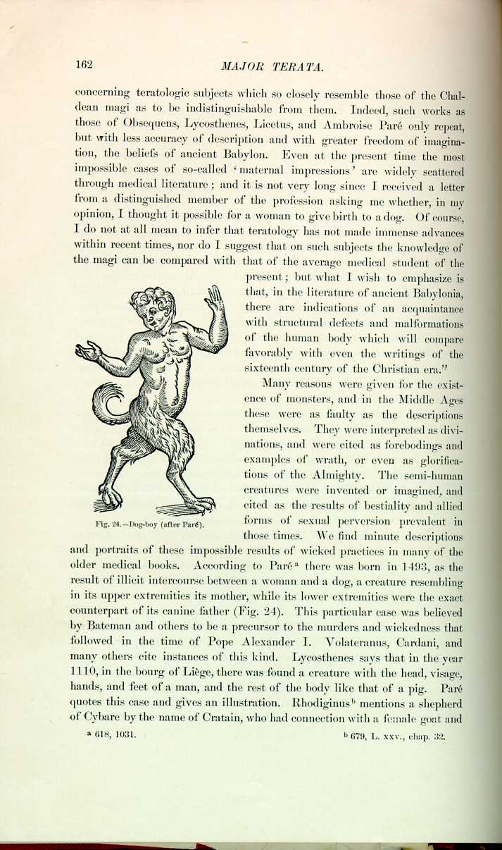
Fig. 24.—Dog-boy (after Paré).
[Description: Drawing of a figure that is part boy, part dog]
We find minute descriptions and portraits of these impossible results of wicked practices in many of the older medical books. According to Paré [5.2] there was born in 1493, as the result of illicit intercourse between a woman and a dog, a creature resembling in its upper extremities its mother, while its lower extremities were the exact counterpart of its canine father (Fig. 24). This particular case was believed by Bateman and others to be a precursor to the murders and wickedness that followed in the time of Pope Alexander I. Volateranus, Cardani, and many others cite instances of this kind. Lycosthenes says that in the year 1110, in the bourg of Liège, there was found a creature with the head, visage, hands, and feet of a man, and the rest of the body like that of a pig. Paré quotes this case and gives an illustration. Rhodiginus [5.3] mentions a shepherd of Cybare by the name of Cratain, who had connection with a female goat and
In the year 1547, at Cracovia, *[191] a very strange monster was born, which lived three days. It had a head shaped like that of a man; a nose long and hooked like an elephant's trunk; the hands and feet looking like the web-foot of a goose; and a tail with a hook on it. It was supposed to be a male, and was looked upon as a result of sodomy. Rueff [5.4] says that the procreation of human beings and beasts is brought about—
(1) By the natural appetite;
(2) By the provocation of nature by delight;
(3) By the attractive virtue of the matrix, which in beasts and women is alike.
Plutarch, in his "Lesser Parallels,'' says that Aristonymus Ephesius, son of Demonstratus, being tired of women, had carnal knowledge with an ass, which in the process of time brought forth a very beautiful child, who became the maid Onoscelin. He also speaks of the origin of the maiden Hippona, or as he calls her, Hippo, as being from the connection of a man with a mare. Aristotle mentions this in his paradoxes, and we know that the patron of horses was Hippona. In Helvetia *[191] was reported the existence of a colt (whose mother had been covered by a bull) that was half horse and half bull. One of the kings of France was supposed to have been presented with a colt with the hinder part of a hart, and which could outrun any horse in the kingdom. Its mother had been covered by a hart.
Writing in 1557, Lycosthenes reports the mythical birth of a serpent by a woman. It is quite possible that some known and classified type of monstrosity was indicated here in vague terms. In 1726 Mary Toft, of Godalming, in Surrey, England, achieved considerable notoriety throughout Surrey, and even over all England, by her extensively circulated statements that she bore rabbits. Even at so late a day as this the credulity of the people was so great that many persons believed in her. The woman was closely watched, and being detected in her maneuvers confessed her fraud. To show the extent of discussion this case called forth, there are no less than nine pamphlets and books in the Surgeon-General's library at Washington devoted exclusively to this case of pretended rabbit-breeding. Hamilton in 1848, and Hard [5.5] in 1884, both report the births in this country of fetal monstrosities with heads which showed marked resemblance to those of dogs. Doubtless many of the older cases of the supposed results of bestiality, if seen to-day, could be readily classified among some of our known forms of monsters. Modern investigation has shown us the sterile results of the connections between man and beast or between beasts of
Before ending these preliminary remarks, there might be mentioned the marine monsters, such as mermaids, sea-serpents, and the like, which from time to time have been reported; even at the present day there are people who devoutly believe that they have seen horrible and impossible demons in the sea. Paré [5.6] describes and pictures a monster, at Rome, on November 3, 1520, with the upper portion of a child apparently about five or six years old, and the lower part and ears of a fish-like animal. He also pictures a sea-devil in the same chapter, together with other gruesome examples of the power of imagination.
Early Teratology.—Besides such cases as the foregoing, we find the medieval writers report likely instances of terata, as, for instance, Rhodiginus, *[679] who speaks of a monster in Italy with two heads and two bodies; Lycosthenes saw a double monster, both components of which slept at the same time; he also says this creature took its food and drink simultaneously in its two mouths. Even Saint Augustine says that he knew of a child born in the Orient who, from the belly up, was in all parts double.
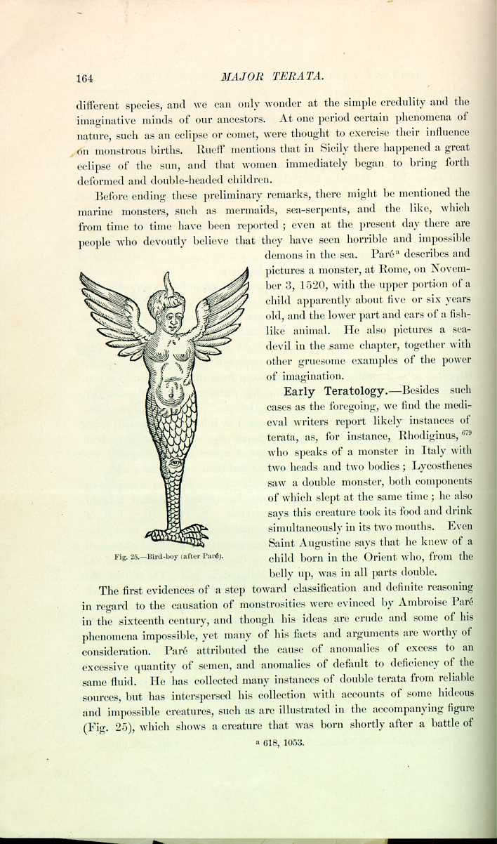
Fig. 25.—Bird-boy (after Paré).
[Description: Drawing of a figure that is half boy, half bird]
The first evidences of a step toward classification and definite reasoning in regard to the causation of monstrosities were evinced by Ambroise Paré in the sixteenth century, and though his ideas are crude and some of his phenomena impossible, yet many of his facts and arguments are worthy of consideration. Paré attributed the cause of anomalies of excess to an excessive quantity of semen, and anomalies of default to deficiency of the same fluid. He has collected many instances of double terata from reliable sources, but has interspersed his collection with accounts of some hideous and impossible creatures, such as are illustrated in the accompanying figure (Fig. 25), which shows a creature that was born shortly after a battle of

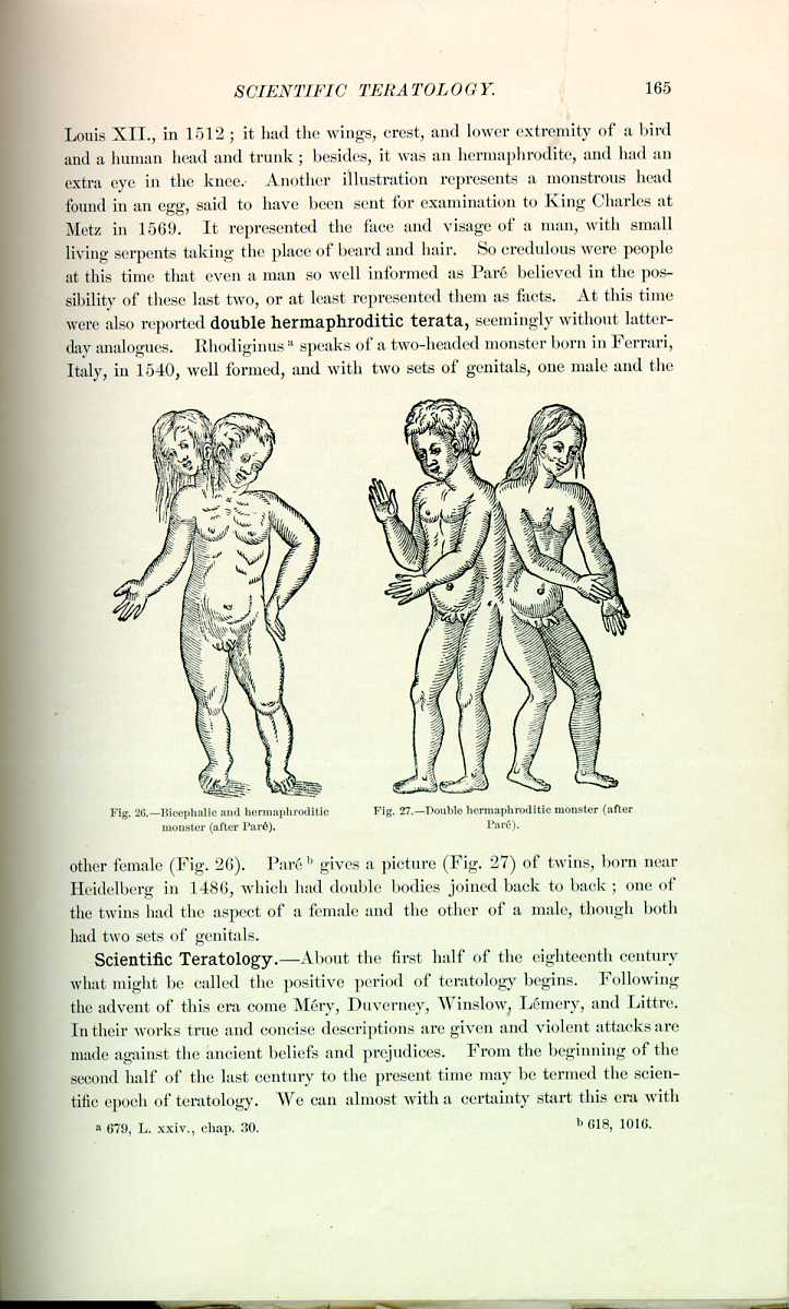
other female (Fig. 26). Paré [5.8] gives a picture (Fig. 27) of twins, born near Heidelberg in 1486, which had double bodies joined back to back; one of the twins had the aspect of a female and the other of a male, though both had two sets of genitals.
Scientific Teratology.—About the first half of the eighteenth century what might be called the positive period of teratology begins. Following the advent of this era come Méry, Duverney, Winslow, Lémery, and Littre. In their works true and concise descriptions are given and violent attacks are made against the ancient beliefs and prejudices. From the beginning of the second half of the last century to the present time may be termed the scientific epoch of teratology. We can almost with a certainty start this era with
Terata are seen in the lower animals and always excite interest. Paré [5.11] gives the history of a sheep with three heads, born in 1577; the central head was larger than the other two, as shown in the accompanying illustration (Fig. 28).
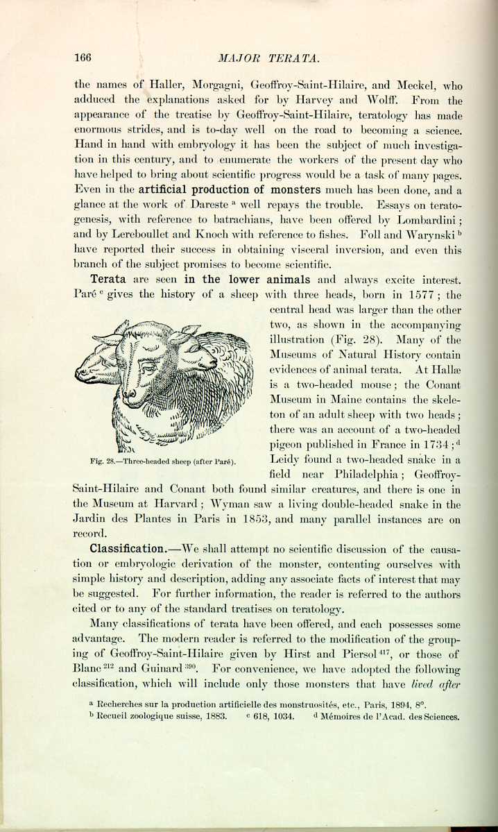
Fig. 28 Three-headed sheep (after Paré).
[Description: Drawing of a three-headed sheep]
Many of the Museums of Natural History contain evidences of animal terata. At Hallæ is a two-headed mouse; the Conant Museum in Maine contains the skeleton of an adult sheep with two heads; there was an account of a two-headed pigeon published in France in 1734; [5.12] Leidy found a two-headed snake in a field near Philadelphia; Geoffroy Saint-Hilaire and Conant both found similar creatures, and there is one in the Museum at Harvard; Wyman saw a living double-headed snake in the Jardin des Plantes in Paris in 1853, and many parallel instances are on record.
Classification.—We shall attempt no scientific discussion of the causation or embryologic derivation of the monster, contenting ourselves with simple history and description, adding any associate facts of interest that may be suggested. For further information, the reader is referred to the authors cited or to any of the standard treatises on teratology.
Many classifications of terata have been offered, and each possesses some advantage. The modern reader is referred to the modification of the grouping of Geoffroy-Saint-Hilaire given by Hirst and Piersol *[417], or those of Blanc *[212] and Guinard *[390]. For convenience, we have adopted the following classification, which will include only those monsters that have lived after
- CLASS 1.—Union of several fetuses.
- CLASS 2.—Union of two distinct fetuses by a connecting band.
- CLASS 3.—Union of two distinct fetuses by an osseous junction of the cranial bones.
- CLASS 4.—Union of two distinct fetuses in which one or more parts are eliminated by the junction.
- CLASS 5.—Fusion of two fetuses by a bony union of the ischii.
- CLASS 6.—Fusion of two fetuses below the umbilicus into a common lower extremity.
- CLASS 7.—Bicephalic monsters.
- CLASS 8.—Parasitic monsters.
- CLASS 9.—Monsters with a single body and double lower extremities.
- CLASS 10.—Diphallic terata.
- CLASS 11.—Fetus in fetu, and dermoid cysts.
- CLASS 12.—Hermaphrodites.
CLASS I.—Triple Monsters.—Haller and Meckel were of the opinion that no cases of triple monsters worthy of credence are on record, and since their time this has been the popular opinion. Surely none have ever lived. Licetus *[486] describes a human monster with two feet and seven heads and as many arms. Bartholinus [5.13] speaks of a three-headed monster who after birth gave vent to horrible cries and expired. Borellus *[841] speaks of a three-headed dog, a veritable Cerberus. Blasius *[214] published an essay on triple monsters in 1677. Bordenave [5.14] is quoted as mentioning a human monster formed of three fetuses, but his description proves clearly that it was only the union of two. Probably the best example of this anomaly that we have was described by Galvagni at Cattania in 1834. This monster had two necks, on one of which was a single head normal in dimensions.
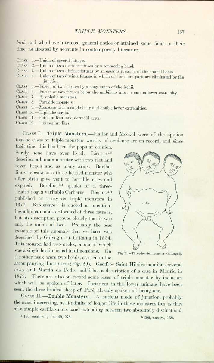
Fig. 29. Three-headed monster (Galvagni).
[Description: Drawing of a boy with three heads]
On the other neck were two heads, as seen in the accompanying illustration (Fig. 29). Geoffroy-Saint-Hilaire mentions several cases, and Martin de Pedro publishes a description of a case in Madrid in 1879. There are also on record some cases of triple monster by inclusion which will be spoken of later. Instances in the lower animals have been seen, the three-headed sheep of Paré, already spoken of, being one.
CLASS II.—Double Monsters.—A curious mode of junction, probably the most interesting, as it admits of longer life in these monstrosities, is that of a simple cartilaginous band extending between two absolutely distinct and
Perhaps the best known of all double monsters were the Siamese twins. They were exhibited all over the globe and had the additional benefit and advertisement of a much mooted discussion as to the advisability of their severance, in which opinions of the leading medical men of all nations were advanced. The literature on these famous brothers is simply stupendous. The amount of material in the Surgeon General's library at Washington would surprise an investigator. A curious volume in this library is a book containing clippings, advertisements, and divers portraits of the twins. It will be impossible to speak at all fully on this subject, but a short history and running review of their lives will be
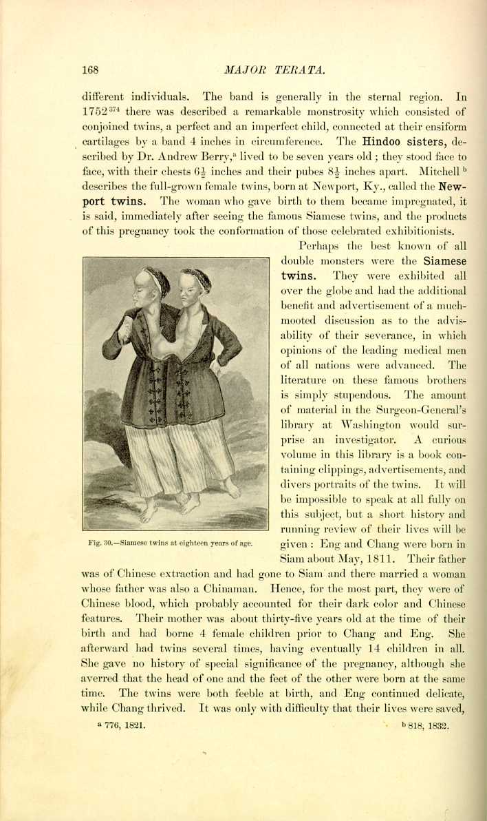
Fig. 30.—Siamese twins at eighteen years of age.
[Description: Drawing of Siamese twins]
given: Eng and Chang were born in Siam about May, 1811. Their father was of Chinese extraction and had gone to Siam and there married a woman whose father was also a Chinaman. Hence, for the most part, they were of Chinese blood, which probably accounted for their dark color and Chinese features. Their mother was about thirty-five years old at the time of their birth and had borne 4 female children prior to Chang and Eng. She afterward had twins several times, having eventually 14 children in all. She gave no history of special significance of the pregnancy, although she averred that the head of one and the feet of the other were born at the same time. The twins were both feeble at birth, and Eng continued delicate, while Chang thrived. It was only with difficulty that their lives were saved,
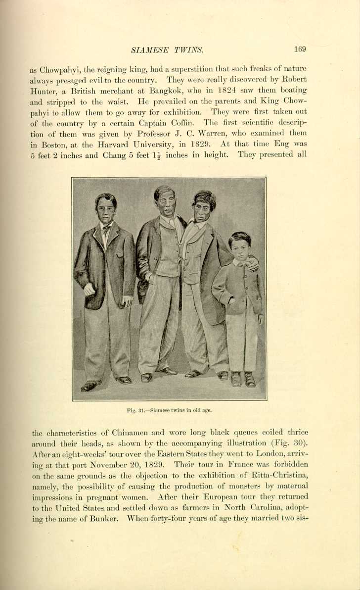
Fig. 31.—Siamese twins in old age.
[Description: Drawing of Siamese twins]
the characteristics of Chinamen and wore long black queues coiled thrice around their heads, as shown by the accompanying illustration (Fig. 30). After an eight-weeks' tour over the Eastern States they went to London, arriving at that port November 20, 1829. Their tour in France was forbidden on the same grounds as the objection to the exhibition of Ritta-Christina, namely, the possibility of causing the production of monsters by maternal impressions in pregnant women. After their European tour they returned to the United States and settled down as farmers in North Carolina, adopting the name of Bunker. When forty-four years of age they married two sisters,
A most pathetic characteristic of these illustrious brothers was the affection and forbearance they showed for each other until shortly before their death. They bore each other's trials and petty maladies with the greatest sympathy, and in this manner rendered their lives far more agreeable than a casual observer would suppose possible. They both became Christians and members or attendants of the Baptist Church.
Figure 31 is a representation of the Siamese twins in old age. On each side of them is a son. The original photograph is in the Mütter Museum, College of Physicians, Philadelphia.
The feasibility of the operation of separating them was discussed by many of the leading men of America, and Thompson, Fergusson, Syme, Sir J. Y. Simpson,
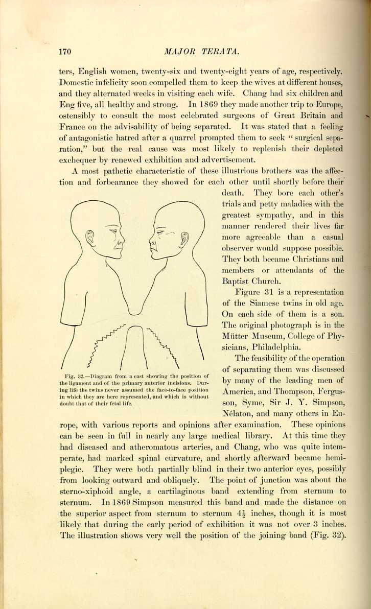
Nélaton, and many others in Europe, with various reports and opinions after examination. These opinions can be seen in full in nearly any large medical library. At this time they had diseased and atheromatous arteries, and Chang, who was quite intemperate, had marked spinal curvature, and shortly afterward became hemiplegic. They were both partially blind in their two anterior eyes, possibly from looking outward and obliquely. The point of junction was about the sterno-siphoid angle, a cartilaginous band extending from sternum to sternum. In 1869 Simpson measured this band and made the distance on the superior aspect from sternum to sternum 4 1/2 inches, though it is most likely that during the early period of exhibition it was not over 3 inches. The illustration shows very well the position of the joining band (Fig. 32).
The twins died on January 17, 1874, and a committee of surgeons from the College of Physicians of Philadelphia, consisting of Doctors Andrews, Allen, and Pancoast, went to North Carolina to perform an autopsy on the body, and, if possible, to secure it. They made a long and most interesting report on the results of their trip to the College. The arteries, as was anticipated, were found to have undergone calcareous degeneration. There was an hepatic connection through the band, and also some interlacing diaphragmatic fibers therein. There was slight vascular intercommunication of the livers and independence of the two peritoneal cavities and the intestines (Fig. 33). The band itself was chiefly a coalescence of the xyphoid cartilages, surrounded by areolar tissue and skin (Fig. 34).
The "Orissa sisters,'' or Radica-Doddica, shown in Europe in 1893, were similar to the Siamese twins in conformation. They were born in

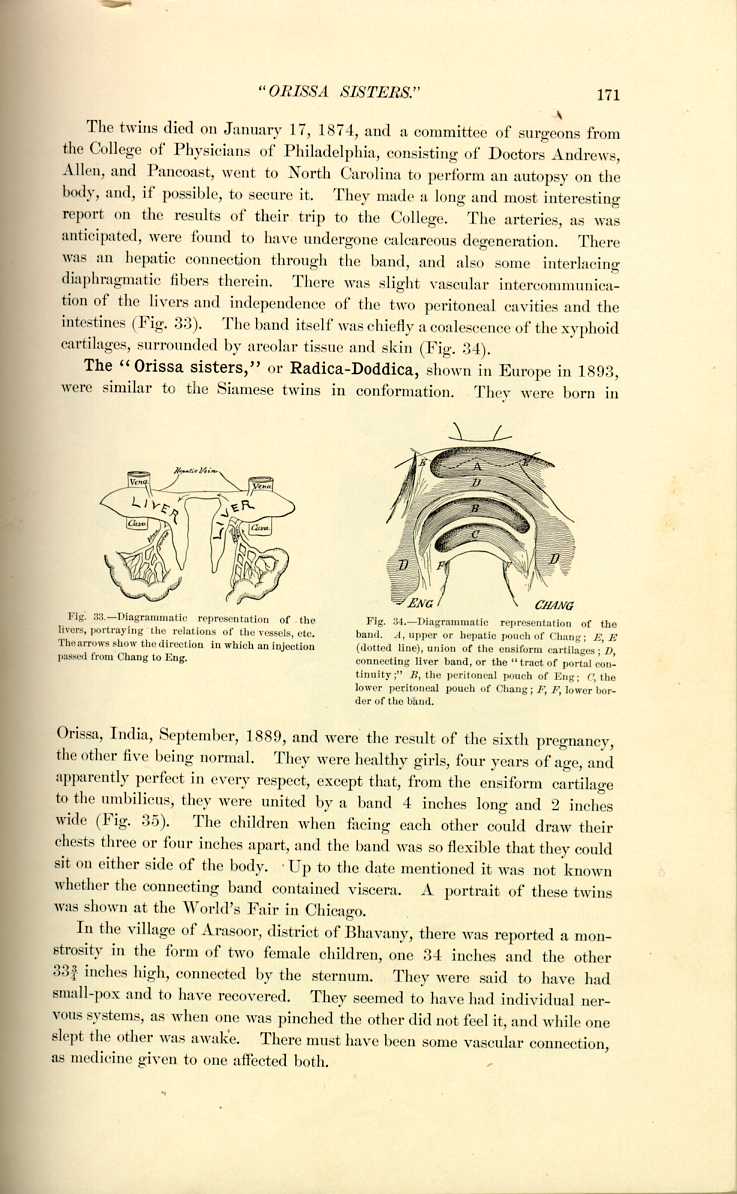
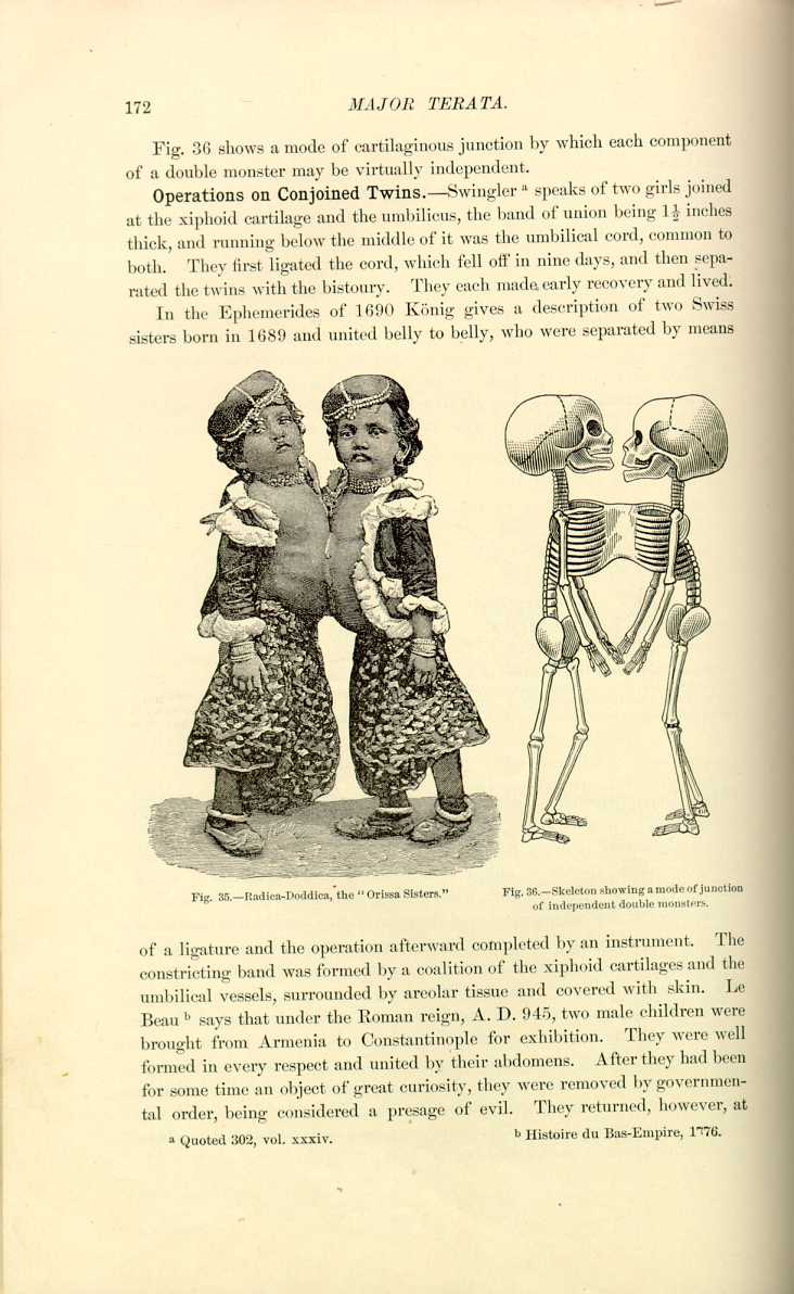
Orissa, India, September, 1889, and were the result of the sixth pregnancy, the other five being normal. They were healthy girls, four years of age, and apparently perfect in every respect, except that, from the ensiform cartilage to the umbilicus, they were united by a band 4 inches long and 2 inches wide (Fig. 35). The children when facing each other could draw their chests three or four inches apart, and the band was so flexible that they could sit on either side of the body. Up to the date mentioned it was not known whether the connecting band contained viscera. A portrait of these twins was shown at the World's Fair in Chicago.
In the village of Arasoor, district of Bhavany, there was reported a monstrosity in the form of two female children, one 34 inches and the other 33 3/4 inches high, connected by the sternum. They were said to have had small-pox and to have recovered. They seemed to have had individual nervous systems, as when one was pinched the other did not feel it, and while one slept the other was awake. There must have been some vascular connection, as medicine given to one affected both.
Operations on Conjoined Twins.—Swingler [5.17] speaks of two girls joined at the xiphoid cartilage and the umbilicus, the band of union being 1 1/2 inches thick, and running below the middle of it was the umbilical cord, common to both. They first ligated the cord, which fell off in nine days, and then separated the twins with the bistoury. They each made early recovery and lived.
In the Ephemerides of 1690 König gives a description of two Swiss sisters born in 1689 and united belly to belly, who were separated by means

Fig. 35.—Radica-Doddica, the "Orissa Sisters.''
[Description: Drawing of the "Orissa Sisters"]
of a ligature and the operation afterward completed by an instrument. The constricting band was formed by a coalition of the xiphoid cartilages and the umbilical vessels, surrounded by areolar tissue and covered with skin. Le Beau [5.18] says that under the Roman reign, A. D. 945, two male children were brought from Armenia to Constantinople for exhibition. They were well formed in every respect and united by their abdomens. After they had been for some time an object of great curiosity, they were removed by governmental order, being considered a presage of evil. They returned, however, at
In 1866 Boehm [5.19] gives an account of Guzenhausen's case of twins who were united sternum to sternum. An operation for separation was performed without accident, but one of the children, already very feeble, died three days after; the other survived. The last attempt at an operation like this was in 1881, when Biaudet and Buginon attempted to separate conjoined sisters (Marie-Adèle) born in Switzerland on June 26th. Unhappily, they were very feeble and life was despaired of when the operation was performed, on October 29th. Adèle died six hours afterward, and Marie died of peritonitis on the next day.
CLASS III.—Those monsters joined by a fusion of some of the cranial bones are sometimes called craniopagi. A very ancient observation of this kind is cited by Geoffroy-Saint-Hilaire. These two girls were born in 1495, and lived to be ten years old. They were normal in every respect, except that they were joined at the forehead, causing them to stand face to face and belly to belly (Fig. 37). When one walked forward, the other was compelled to walk backward; their noses almost touched, and their eyes were directed laterally. At the death of one an attempt to separate the other from the cadaver was made, but it was unsuccessful, the second soon dying;

Fig. 37.—Craniopagus (after Paré).
[Description: Drawing of women joined by fusion of cranial bones]
the operation necessitated opening the cranium and parting the meninges. Bateman *[191] said that in 1501 there was living an instance of double female twins, joined at the forehead. This case was said to have been caused in the following manner: Two women, one of whom was pregnant with the twins at the time, were engaged in an earnest conversation, when a third, coming up behind them, knocked their heads together with a sharp blow. Bateman describes the death of one of the twins and its excision from the other, who died subsequently, evidently of septic infection. There is a possibility that this is merely a duplication of the account of the preceding case with a slight anachronism as to the time of death.
At a foundling hospital in St. Petersburg [5.20] there were born two living girls, in good health, joined by the heads. They were so united that the nose of one, if prolonged, would strike the ear of the other; they had perfectly independent existences, but their vascular systems had evident connection.
CLASS IV.—The next class to be considered is that in which the individuals are separate and well formed, except that the point of fusion is a common part, eliminating their individual components in this location. The pygopagous twins belong in this section. According to Bateman, *[191] twins were born in 1493 at Rome joined back to back, and survived their birth. The same authority speaks of a female child who was born with "2 bellies, 4 arms, 4 legs, 2 heads, and 2 sets of privates, and was exhibited throughout Italy for gain's sake.'' The "Biddenden Maids'' were born in Biddenden, Kent, in 1100. [5.23] Their names were Mary and Eliza Chulkhurst, and their parents were fairly well-to-do people. They were supposed to have been united at the hips and the shoulders, and lived until 1134. At the death of one it was proposed to separate them, but the remaining sister refused, saying, "As we came together, we will also go together,'' and, after about six hours of this Mezentian existence, they died. They bequeathed to the church-wardens of the parish and their successors land to the extent of 20 acres, at the present time bringing a rental of about $155.00 annually, with the instructions that the money was to be spent in the distribution of cakes
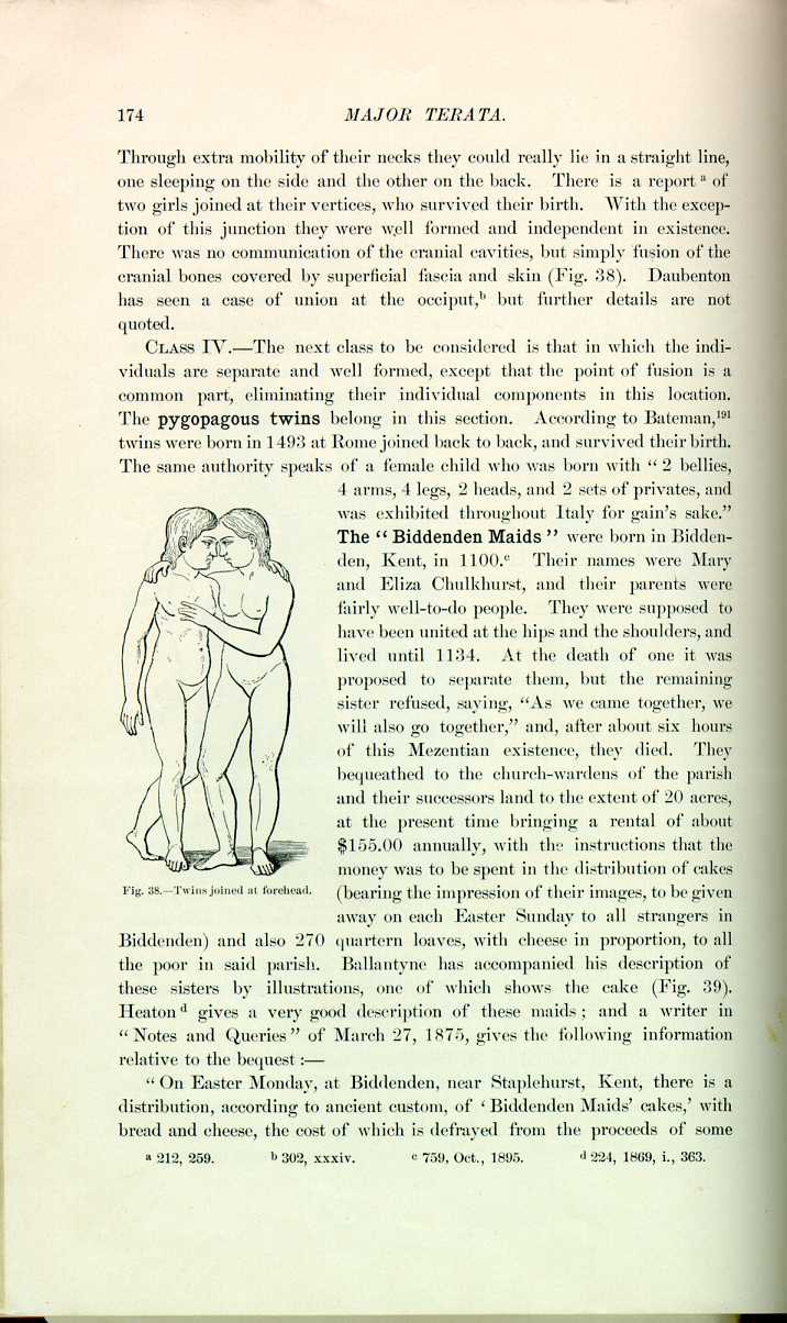
Fig. 38—Twins joined at forehead.
[Description: Drawing of twins joined at the forehead]
(bearing the impression of their images, to be given away on each Easter Sunday to all strangers in Biddenden) and also 270 quartern loaves, with cheese in proportion, to all the poor in said parish. Ballantyne has accompanied his description of these sisters by illustrations, one of which shows the cake (Fig. 39). Heaton [5.24] gives a very good description of these maids; and a writer in "Notes and Queries'' of March 27, 1875, gives the following information relative to the bequest:—
"On Easter Monday, at Biddenden, near Staplehurst, Kent, there is a distribution, according to ancient custom, of `Biddenden Maids' cakes,' with bread and cheese, the cost of which is defrayed from the proceeds of some
Ballantyne *[759] has summed up about all there is to be said on this national monstrosity, and his discussion of the case from its historic as well as teratologic standpoint is so excellent that
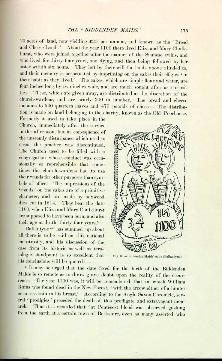
Fig. 39.—Biddenden Maids' cake (Ballantyne).
[Description: Drawing of the Biddenden Maids' cake]
his conclusions will be quoted—
"It may be urged that the date fixed for the birth of the Biddenden Maids is so remote as to throw grave doubt upon the reality of the occurrence. The year 1100 was, it will be remembered, that in which William Rufus was found dead in the New Forest, `with the arrow either of a hunter or an assassin in his breast.' According to the Anglo-Saxon Chronicle, several `prodigies' preceded the death of this profligate and extravagant monarch. Thus it is recorded that `at Pentecost blood was observed gushing from the earth at a certain town of Berkshire, even as many asserted who
"There is, however, one real difficulty in accepting the story handed down to us as authentic,—the nature of the teratologic phenomenon itself. All the records agree in stating that the Maids were joined together at the shoulders and hips, and the impression on the cakes and the pictures on the `broadsides' show this peculiar mode of union, and represent the bodies as quite separate in the space between the above-named points. The Maids are shown with four feet and two arms, the right and left respectively, whilst the other arms (left and right) are fused together at the shoulder according to one illustration, and a little above the elbow according to another. Now, although it is not safe to say that such an anomaly is impossible, I do not know of any case of this peculiar mode of union; but it may be that, as Prof. A. R. Simpson has suggested, the Maids had four separate arms, and were in the habit of going about with their contiguous arms round each other's necks, and that this gave rise to the notion that these limbs were united. If this be so, then the teratologic difficulty is removed, for the case becomes perfectly comparable with the well-known but rare type of double terata known as the pygopagous twins, which is placed by Taruffi with that of the ischiopagous twins in the group dicephalus lecanopagus. Similar instances, which are well known to students of teratology, are the Hungarian sisters (Helen and Judith), the North Carolina twins (Millie and Christine), and the Bohemian twins (Rosalie and Josepha Blazek). The interspace between the thoraces may, however, have simply been the addition
"Pygopagous twins are fetuses united together in the region of the nates and having each its own pelvis. In the recorded cases the union has been usually between the sacra and coccyges, and has been either osseous or (more rarely) ligamentous. Sometimes the point of junction was the middle line posteriorly, at other times it was rather a posterolateral union; and it is probable that in the Biddenden Maids it was of the latter kind; and it is likely, from the proposal made to separate the sisters after the death of one, that it was ligamentous in nature.
"If it be granted that the Biddenden Maids were pygopagous twins, a study of the histories of other recorded cases of this monstrosity serves to demonstrate many common characters. Thus, of the 8 cases which Taruffi has collected, in 7 the twins were female; and if to these we add the sisters Rosalie and Josepha Blazek and the Maids, we have 10 cases, of which 9 were girls. Again, several of the pygopagous twins, of whom there are scientific records, survived birth and lived for a number of years, and thus resembled the Biddenden terata. Helen and Judith, for instance, were twenty-three years old at death; and the North Carolina twins, although born in 1851, are still alive. There is, therefore, nothing inherently improbable in the statement that the Biddenden Maids lived for thirty-four years. With regard also to the truth of the record that the one Maid survived her sister for six hours, there is confirmatory evidence from scientifically observed instances, for Joly and Peyrat (Bull. de l'Acad. Méd., iii., pp. 51 and 383, 1874) state that in the case seen by them the one infant lived ten hours after the death of the other. It is impossible to make any statement with regard to the internal structure of the Maids or to the characters of their genital organs, for there is absolutely no information forthcoming upon these points. It may simply be said, in conclusion, that the phenomenon of Biddenden is interesting not only on account of the curious bequest which arose out of it, but also because it was an instance of a very rare teratologic type, occurring at a very early period in our national history.''
Possibly the most famous example of twins of this type were Helen and Judith, the Hungarian sisters, born in 1701 at Szony, in Hungary. They were the objects of great curiosity, and were shown successively in Holland, Germany, Italy, France, England, and Poland. At the age of nine they were placed in a convent, where they died almost simultaneously in their twenty-second year. During their travels all over Europe they were examined by many prominent physiologists, psychologists, and naturalists; Pope and several minor poets have celebrated their existence in verse; Buffon speaks of them in his "Natural History,'' and all the works on teratology for a century or more have mentioned them. A description of them can be best
That naught their bodies can divide, no power beneath the sun.
The town of Szoenii gave them birth, hard by far-famed Komorn,
Which noble fort may all the arts of Turkish sultans scorn.
Lucina, woman's gentle friend, did Helen first receive;
And Judith, when three hours had passed, her mother's womb did leave.
One urine passage serves for both;—one anus, so they tell;
The other parts their numbers keep, and serve their owners well.
Their parents poor did send them forth, the world to travel through,
That this great wonder of the age should not be hid from view.
The inner parts concealed do lie hid from our eyes, alas!
But all the body here you view erect in solid brass.
They were joined back to back in the lumbar region, and had all their parts separate except the anus between the right thigh of Helen and the left of Judith and a single vulva. Helen was the larger, better looking, the more active, and the more intelligent. Judith at the age of six became hemiplegic, and afterward was rather delicate and depressed. They menstruated at sixteen and continued with regularity, although one began before the other. They had a mutual affection, and did all in their power to alleviate the circumstances of their sad position. Judith died of cerebral and pulmonary affections, and Helen, who previously enjoyed good health, soon after her sister's first indisposition suddenly sank into a state of collapse, although preserving her mental faculties, and expired almost immediately after her sister. They had measles and small-pox simultaneously, but were affected in different degree by the maladies. The emotions, inclinations, and appetites were
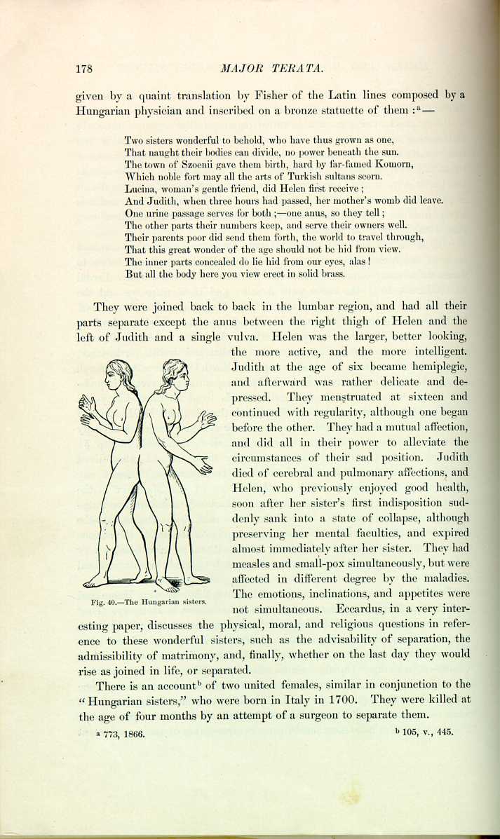
Fig. 40.—The Hungarian sisters.
[Description: Drawing of the Hungarian sisters]
not simultaneous. Eccardus, in a very interesting paper, discusses the physical, moral, and religious questions in reference to these wonderful sisters, such as the advisability of separation, the admissibility of matrimony, and, finally, whether on the last day they would rise as joined in life, or separated.
There is an account [5.26] of two united females, similar in conjunction to the "Hungarian sisters,'' who were born in Italy in 1700. They were killed at the age of four months by an attempt of a surgeon to separate them.
Pancoast [5.27] gives an extensive report of Millie-Christine, who had been extensively exhibited in Europe and the United States. They were born of slave parents in Columbus County, N. C., July 11, 1851; the mother, who had borne 8 children before, was a stout negress of thirty-two, with a large pelvis. The presentation was first by the stomach and afterward by the breech. These twins were united at the sacra by a cartilaginous or possibly osseous union. They were exhibited in Paris in 1873, and provoked as much discussion there as in the United States. Physically, Millie was the weaker, but had the stronger will and the dominating spirit. They menstruated regularly from the age of thirteen. One from long habit yielded instinctively to the other's movements, thus preserving the necessary harmony. They ate separately, had distinct thoughts, and carried on distinct conversations at the same time. They experienced hunger and thirst generally simultaneously, and defecated and urinated nearly at the same times. One, in tranquil sleep, would be wakened by a call of nature of the other. Common sensibility was experienced near the location of union. They were intelligent and agreeable
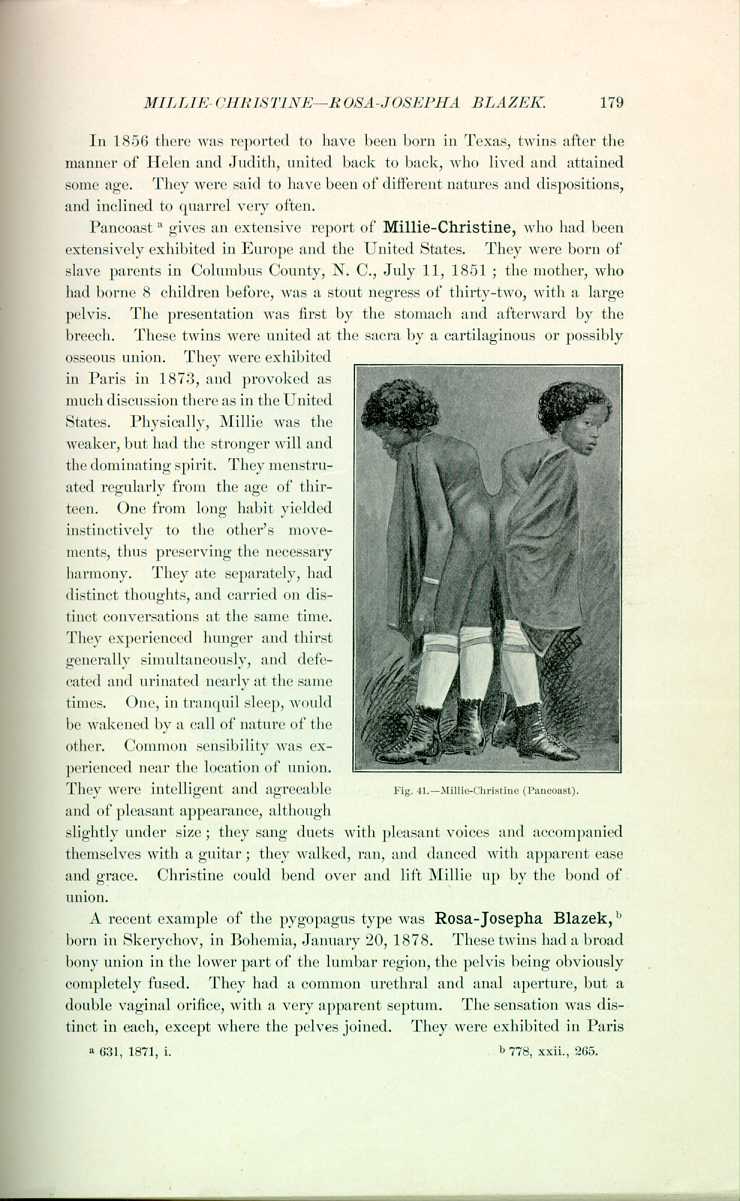
Fig. 41.—Mille-Christine (Pancoast).
[Description: Drawing of Millie-Christine]
and of pleasant appearance, although slightly under size; they sang duets with pleasant voices and accompanied themselves with a guitar; they walked, ran, and danced with apparent ease and grace. Christine could bend over and lift Millie up by the bond of union.
A recent example of the pygopagus type was Rosa-Josepha Blazek, [5.28] born in Skerychov, in Bohemia, January 20, 1878. These twins had a broad bony union in the lower part of the lumbar region, the pelvis being obviously completely fused. They had a common urethral and anal aperture, but a double vaginal orifice, with a very apparent septum. The sensation was distinct in each, except where the pelves joined. They were exhibited in Paris
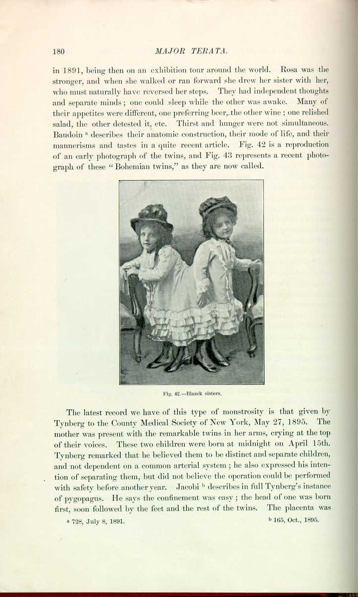
Fig. 42.—Blazek sisters.
[Description: Reproduction of a photograph of the Blazek twins]
The latest record we have of this type of monstrosity is that given by Tynberg to the County Medical Society of New York, May 27, 1895. The mother was present with the remarkable twins in her arms, crying at the top of their voices. These two children were born at midnight on April 15th. Tynberg remarked that he believed them to be distinct and separate children, and not dependent on a common arterial system; he also expressed his intention of separating them, but did not believe the operation could be performed with safety before another year. Jacobi [5.30] describes in full Tynberg's instance of pygopagus. He says the confinement was easy; the head of one was born first, soon followed by the feet and the rest of the twins. The placenta was
CLASS V.—While instances of ischiopagi are quite numerous, few have attained any age, and, necessarily, little notoriety. Paré [5.31] speaks of twins
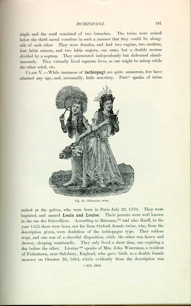
Fig. 43.—Bohemian twins.
[Description: Drawing of a photograph of the Bohemian twins]
united at the pelves, who were born in Paris July 20, 1570. They were baptized, and named Louis and Louise. Their parents were well known in the rue des Gravelliers. According to Bateman, *[191] and also Rueff, in the year 1552 there were born, not far from Oxford, female twins, who, from the description given, were doubtless of the ischiopagus type. They seldom wept, and one was of a cheerful disposition, while the other was heavy and drowsy, sleeping continually. They only lived a short time, one expiring a day before the other. Licetus *[486] speaks of Mrs. John Waterman, a resident of Fishertown, near Salisbury, England, who gave birth to a double female monster on October 26, 1664, which evidently from the description was
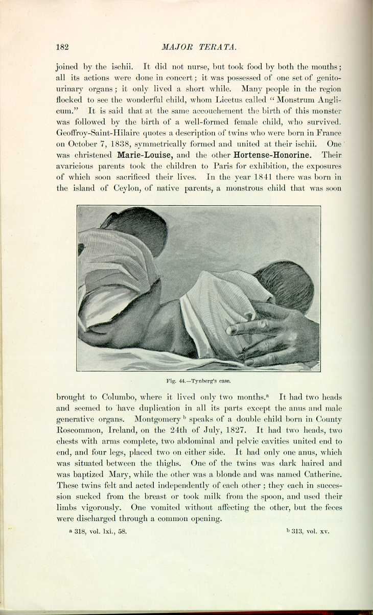
Fig. 44.—Tynberg's case.
[Description: Drawing of Tynberg's twins]
brought to Columbo, where it lived only two months. [5.32] It had two heads and seemed to have duplication in all its parts except the anus and male generative organs. Montgomery [5.33] speaks of a double child born in County Roscommon, Ireland, on the 24th of July, 1827. It had two heads, two chests with arms complete, two abdominal and pelvic cavities united end to end, and four legs, placed two on either side. It had only one anus, which was situated between the thighs. One of the twins was dark haired and was baptized Mary, while the other was a blonde and was named Catherine. These twins felt and acted independently of each other; they each in succession sucked from the breast or took milk from the spoon, and used their limbs vigorously. One vomited without affecting the other, but the feces were discharged through a common opening.
Huff [5.36] details a description of the "Jones twins,'' born on June 24, 1889, in Tipton County, Indiana, whose spinal columns were in apposition at the lower end. The labor, of less than two hours' duration, was completed
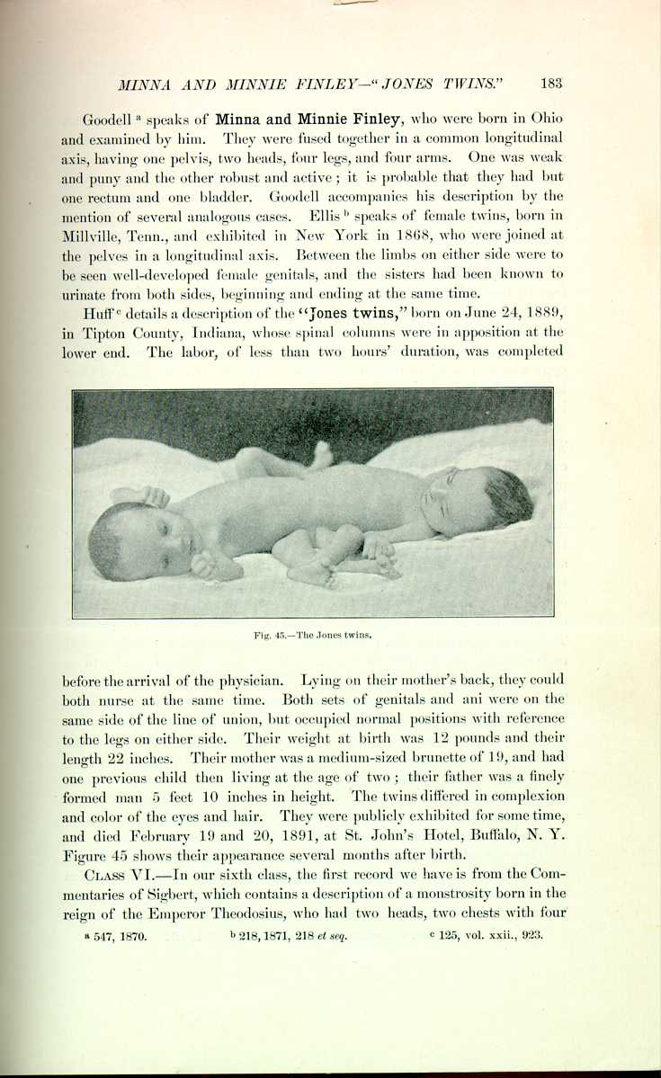
Fig. 45.—The Jones twins.
[Description: Photograph of the Jones twins]
before the arrival of the physician. Lying on their mother's back, they could both nurse at the same time. Both sets of genitals and ani were on the same side of the line of union, but occupied normal positions with reference to the legs on either side. Their weight at birth was 12 pounds and their length 22 inches. Their mother was a medium-sized brunette of 19, and had one previous child then living at the age of two; their father was a finely formed man 5 feet 10 inches in height. The twins differed in complexion and color of the eyes and hair. They were publicly exhibited for some time, and died February 19 and 20, 1891, at St. John's Hotel, Buffalo, N. Y. Figure 45 shows their appearance several months after birth.
CLASS VI.—In our sixth class, the first record we have is from the Commentaries of Sigbert, which contains a description of a monstrosity born in the reign of the Emperor Theodosius, who had two heads, two chests with four
Roger of Wendover *[689] says that in Lesser Brittany and Normandy, in 1062, there was seen a female monster, consisting of two women joined about the umbilicus and fused into a single lower extremity. They took their food by two mouths but expelled it at a single orifice. At one time, one of the women laughed, feasted, and talked, while the other wept, fasted, and kept a religious silence. The account relates how one of them died, and the survivor bore her dead sister about for three years before she was overcome by the oppression and stench of the cadaver. Batemen *[191] describes the birth of a boy in 1529, who had two heads, four ears, four arms, but only two thighs and two legs. Buchanan [5.37] speaks at length of the famous "Scottish Brothers,'' who were the cynosure of the eyes of the Court of James III. of Scotland. This monster consisted of two men, ordinary in appearance in the superior extremities, whose trunks fused into a single lower extremity. The King took diligent care of their education, and they became proficient in music, languages, and other court accomplishments. Between them they would carry on animated conversations, sometimes merging into curious debates, followed by blows. Above the point of union they had no synchronous sensations, while below, sensation was common to both. This monster lived twenty-eight years, surviving the royal patron, who died June, 1488. One of the brothers died some days before the other, and the survivor, after carrying about his dead brother, succumbed to "infection from putrescence.'' There was reported to have been born *[191] in Switzerland a double headed male monster, who in 1538, at the age of thirty, was possessed of a beard on each face, the two bodies fused at the umbilicus into a single lower extremity. These two twins resembled one another in contour and countenance. They were so joined that at rest they looked upon one another. They had a single wife, with whom they were said to have lived in harmony. In the Gentleman's Magazine about one hundred and fifty years since there was given the portrait and description of a double woman, who was exhibited all over the large cities of Europe. Little can be ascertained anatomically of her construction, with the exception that it was stated that she had two heads, two necks, four arms, two legs, one pelvis, and one set of pelvic organs.
The most celebrated monster of this type was Ritta-Christina(Figs. 46 and 47), who was born in Sassari, in Sardinia, March 23, 1829. These twins were the result of the ninth confinement of their mother, a woman of thirty-two. Their superior extremities were double, but they joined in a common trunk

Fig. 46.—Skeleton of Ritta-Christina.
[Description: Drawing of skeleton of Ritta-Christina]
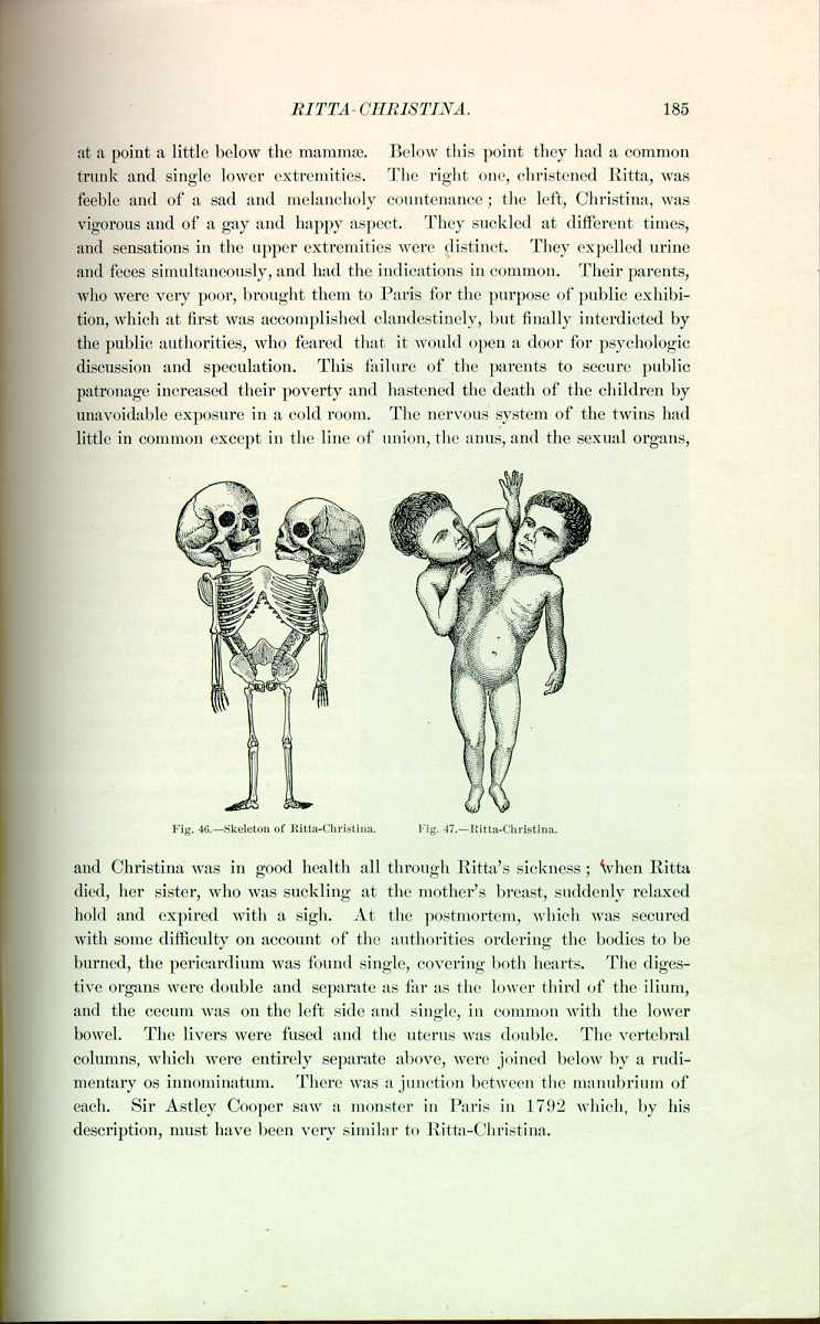
Fig. 47.—Ritta-Christina.
[Description: Drawing of Ritta-Christina]
and Christina was in good health all through Ritta's sickness; when Ritta died, her sister, who was suckling at the mother's breast, suddenly relaxed hold and expired with a sigh. At the postmortem, which was secured with some difficulty on account of the authorities ordering the bodies to be burned, the pericardium was found single, covering both hearts. The digestive organs were double and separate as far as the lower third of the ilium, and the cecum was on the left side and single, in common with the lower bowel. The livers were fused and the uterus was double. The vertebral columns, which were entirely separate above, were joined below by a rudimentary os innorminatum. There was a junction between the manubrium of each. Sir Astley Cooper saw a monster in Paris in 1792 which, by his description, must have been very similar to Ritta-Christina.
McCallum [5.38] saw two female children in Montreal in 1878 named Marie-Rosa Drouin. They formed a right angle with their single trunk, which commenced at the lower part of the thorax of each. They had a single genital fissure and the external organs of generation of a female. A little over three inches from the anus was a rudimentary limb with a movable articulation; it measured five inches in length and tapered to a fine point, being furnished with a distinct nail, and it contracted strongly to irritation. Marie, the left child, was of fair complexion and more strongly
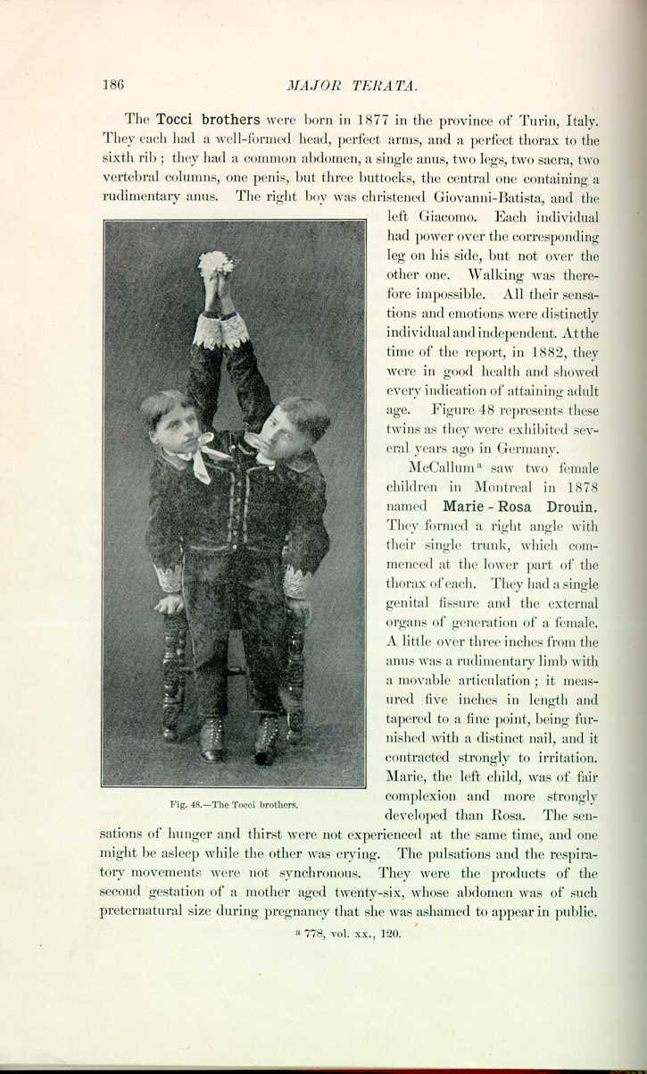
Fig, 48.—The Tocci brothers.
[Description: Photograph of the Tocci brothers]
developed than Rosa. The sensations of hunger and thirst were not experienced at the same time, and one might be asleep while the other was crying. The pulsations and the respiratory movements were not synchronous. They were the products of the second gestation of a mother aged twenty-six, whose abdomen was of such preternatural size during pregnancy that she was ashamed to appear in public.
CLASS VII.—There are many instances of bicephalic monsters on record. Paré [5.39] mentions and gives an illustration of a female apparently single in conformation, with the exception of having two heads and two necks. The Ephemerides, Haller, Schenck, and Archenholz cite examples, and there is an old account [5.40] of a double-headed child, each of whose heads were baptized, one called Martha and the other Mary. One was of a gay and the other a sad visage, and both heads received nourishment; they only lived a couple of days. There is another similar record of a Milanese girl who had two heads, but was in all other respects single, with the exception that after death she was found to have had two stomachs. Besse mentions a Bavarian woman of twenty-six with two heads, one of which was comely and the other extremely ugly; Batemen *[191] quotes what is apparently the same case—a woman in Bavaria in 1541 with two heads, one of which was deformed, who begged from door to door, and who by reason of the influence of pregnant women was given her expenses to leave the country.
A more common occurrence of this type is that in which there is fusion of the two heads. Moreau [5.41] speaks of a monster in Spain which was shown from town to town. Its heads were fused; it had two mouths and two noses; in each face an eye well conformed and placed above the nose; there was a third eye in the middle of the forehead common to both heads; the third eye was of primitive development and had two pupils. Each face was well formed and had its own chin. Buffon mentions a cat, the exact analogue of Moreau's case. Sutton [5.42] speaks of a photograph sent to Sir James Paget in 1856 by William Budd of Bristol. This portrays a living child with a supernumerary head, which had mouth, nose, eyes, and a brain of its own (Fig. 49). The eyelids were abortive, and as there was no orbital cavity the eyes stood out in the form of naked globes on the forehead. When born, the corneas of both heads were transparent, but then became opaque from exposure. The brain of the supernumerary head was quite visible from without, and was covered by a membrane beginning to slough. On the right side of the head was a rudimentary external ear. The nurse said that when the child sucked some milk regurgitated through the supernumerary mouth. The great physiologic interest in this case lies in the fact that every movement and every act of the natural face was simultaneously repeated by the supernumerary face in a perfectly consensual manner, i. e., when the natural mouth sucked, the second mouth sucked; when the natural face cried, yawned, or sneezed, the second face did likewise; and the eyes of the two heads moved in unison. The fate of the child is not known.
Home [5.43] speaks of a child born in Bengal with a most peculiar fusion of
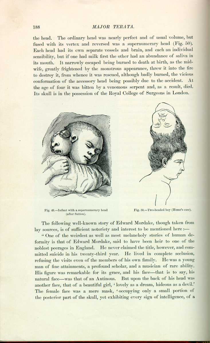

Fig. 50.—Two-headed boy (Home's case).
[Description: Drawing of a two-headed boy]
The following well-known story of Edward Mordake, though taken from lay sources, is of sufficient notoriety and interest to be mentioned here:—
"One of the weirdest as well as most melancholy stories of human deformity is that of Edward Mordake, said to have been heir to one of the noblest peerages in England. He never claimed the title, however, and committed suicide in his twenty-third year. He lived in complete seclusion, refusing the visits even of the members of his own family. He was a young man of fine attainments, a profound scholar, and a musician of rare ability. His figure was remarkable for its grace, and his face—that is to say, his natural face—was that of an Antinous. But upon the back of his head was another face, that of a beautiful girl, `lovely as a dream, hideous as a devil.' The female face was a mere mask, `occupying only a small portion of the posterior part of the skull, yet exhibiting every sign of intelligence, of a
A most curious case was that of a Fellah woman [5.44] who was delivered at Alexandria of a bicephalic monster of apparently eight months' pregnancy. This creature, which was born dead, had one head white and the other black the change of color commencing at the neck of the black head. The bizarre head was of negro conformation and fully developed, and the colored skin was found to be due to the existence of pigment similar to that found in the black race. The husband of the woman had a light brown skin, like an ordinary Fellah man, and it was ascertained that there were some negro laborers in port during the woman's pregnancy; but no definite information as to her relations with them could be established, and whether this was a case of maternal impression or superfetation can only be a matter of conjecture.
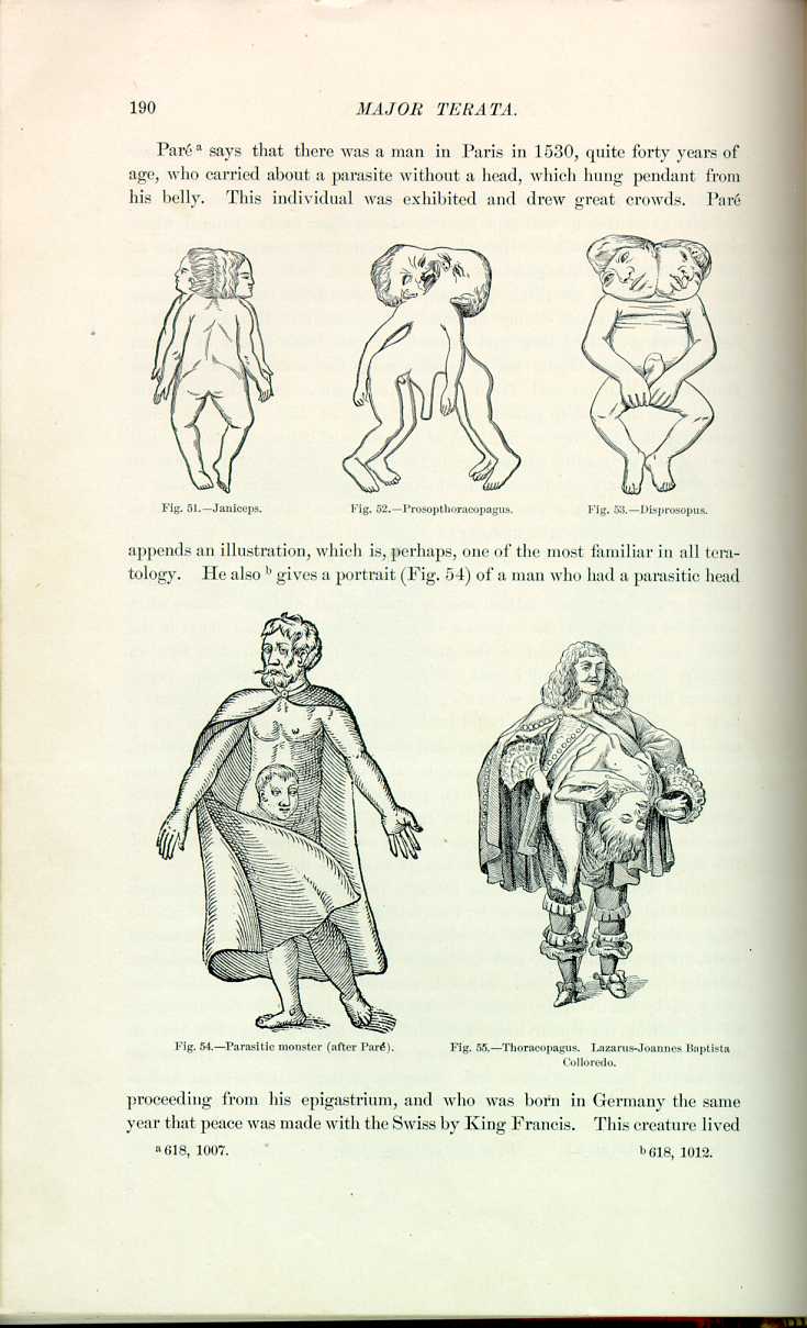
Fantastic monsters, such as acephalon, paracephalon, cyclops, pseudencephalon, and the janiceps (Fig. 51), prosopthoracopagus (Fig. 52), disprosopus (Fig. 53), etc., although full of interest, will not be discussed here, as none are ever viable for any length of time, and the declared intention of this chapter is to include only those beings who have lived.
CLASS VIII.—The next class includes the parasitic terata, monsters that consist of one perfect body, complete in every respect, but from the neighborhood of whose umbilicus depends some important portion of a second body. Paré, Benivenius, and Columbus describe adults with acephalous monsters attached to them. Schenck mentions 13 cases, 3 of which were observed by him. Aldrovandus *[116] shows 3 illustrations under the name of "monstrum bicorpum monocephalon.'' Bustorf [5.45] speaks of a case in which the nates and lower extremities of one body proceeded out of the abdomen of the other, which was otherwise perfect. Reichel and Anderson [5.46] mention a living parasitic monster, the inferior trunk of one body proceeding from the pectoral region of the other.

Fig. 51.—Janiceps.
[Description: Drawing of janiceps]

Fig. 62.—Prosopthoracopagus.
[Description: Drawing of prosopthoracopagus]

Fig. 53.—Disprosopus.
[Description: Drawing of disprosopus]
appends an illustration, which is, perhaps, one of the most familiar in all teratology. He also [5.48] gives a portrait (Fig. 54) of a man who had a parasitic head

Fig. 54.—Parasitic monster (after Paré).
[Description: Drawing of parasitic head on body]
proceeding from his epigastrium, and who was born in Germany the same year that peace was made with the Swiss by King Francis. This creature lived
In 1742, during the Ambassadorship of the Marquis de l'Hôpital at Naples, he saw in that city an aged man, well conformed, with the exception that, like the little girl of Winslow, he had the inferior extremities of a male child growing from his epigastric region. Haller and Meckel have also observed cases like this. Bordat described before the Royal Institute of France, August, 1826, a Chinaman, twenty-one years of age, who had an acephalous fetus attached to the surface of his breast (possibly "A-ke'').
Dickinson [5.50] describes a wonderful child five years old, who, by an extraordinary freak of nature, was an amalgamation of two children. From the body of an otherwise perfectly formed child was a supernumerary head protruding
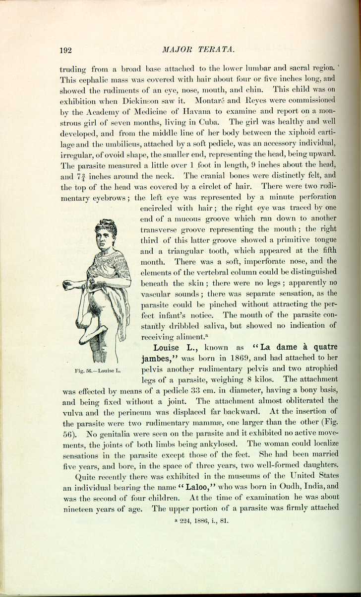
Fig. 56.—Louise L.
[Description: Drawing of Louise L.]
Louise L., known as "La dame à quatre jambes,'' was born in 1869, and had attached to her pelvis another rudimentary pelvis and two atrophied legs of a parasite, weighing 8 kilos. The attachment was effected by means of a pedicle 33 cm. in diameter, having a bony basis, and being fixed without a joint. The attachment almost obliterated the vulva and the perineum was displaced far backward. At the insertion of the parasite were two rudimentary mammæ, one larger than the other (Fig. 56). No genitalia were seen on the parasite and it exhibited no active movements, the joints of both limbs being ankylosed. The woman could localize sensations in the parasite except those of the feet. She had been married five years, and bore, in the space of three years, two well-formed daughters.
Quite recently there was exhibited in the museums of the United States an individual bearing the name "Laloo,'' who was born in Oudh, India, and was the second of four children. At the time of examination he was about nineteen years of age. The upper portion of a parasite was firmly attached
CLASS IX.—Of "Lusus naturæ'' none is more curious than that of duplication of the lower extremities. Paré [5.52] says that on January 9, 1529, there was living in Germany a male infant having four legs and four arms. In Paris, at the Académie des Sciences, on September 6, 1830, there was presented by Madame Hen, a midwife, a living male child with four legs, the anus being nearly below the middle of the third buttock; and the scrotum
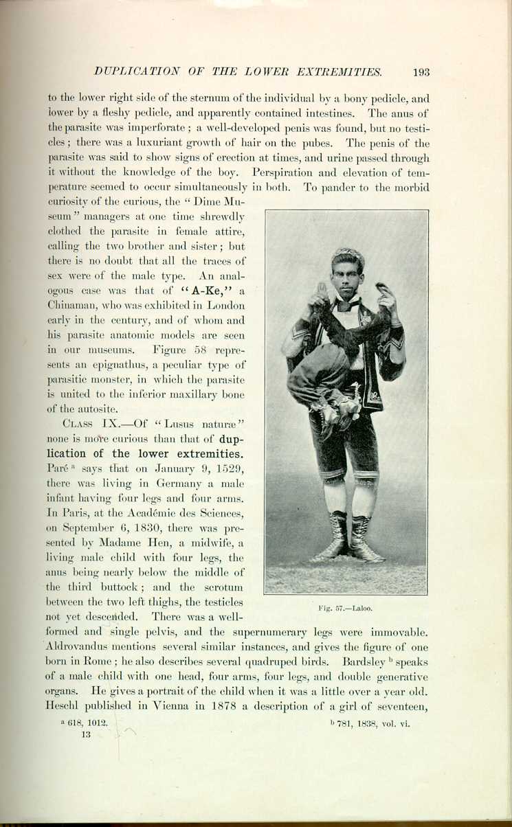
Fig. 57.—Laloo.
[Description: Photograph of Laloo]
between the two left thighs, the testicles not yet descended. There was a well-formed and single pelvis, and the supernumerary legs were immovable. Aldrovandus mentions several similar instances, and gives the figure of one born in Rome; he also describes several quadruped birds. Bardsley [5.53] speaks of a male child with one head, four arms, four legs, and double generative organs. He gives a portrait of the child when it was a little over a year old. Heschl published in Vienna in 1878 a description of a girl of seventeen,
Wells [5.54] describes Mrs. B., aged twenty, still alive and healthy (Fig. 59). The duplication in this case begins just above the waist, the spinal column dividing at the third lumbar vertebra, below this point everything being double. Micturition and defecation occur at different times, but menstruation occurs simultaneously. She was married at nineteen, and became pregnant a year later on the left side, but abortion was induced at the fourth month on account of persistent nausea and the expectation of impossible delivery. Whaley, [5.55] in speaking of this case, said Mrs. B. utilized her outside legs for walking; he also remarks that when he informed her that she was pregnant on the left side she replied, "I think you are mistaken; if it had been on my right side I would come nearer believing it;''—and after further questioning he found, from the patient's observation, that her right genitals were almost invariably used for coitus. Bechlinger of Para, Brazil, [5.56] describes a woman of twenty-five, a native of Martinique, whose father was French and mother a quadroon, who had a modified duplication of the lower body. There was a third leg attached to a continuation of the processus coceygeus of the sacrum, and in addition to well developed mammæ regularly situated,
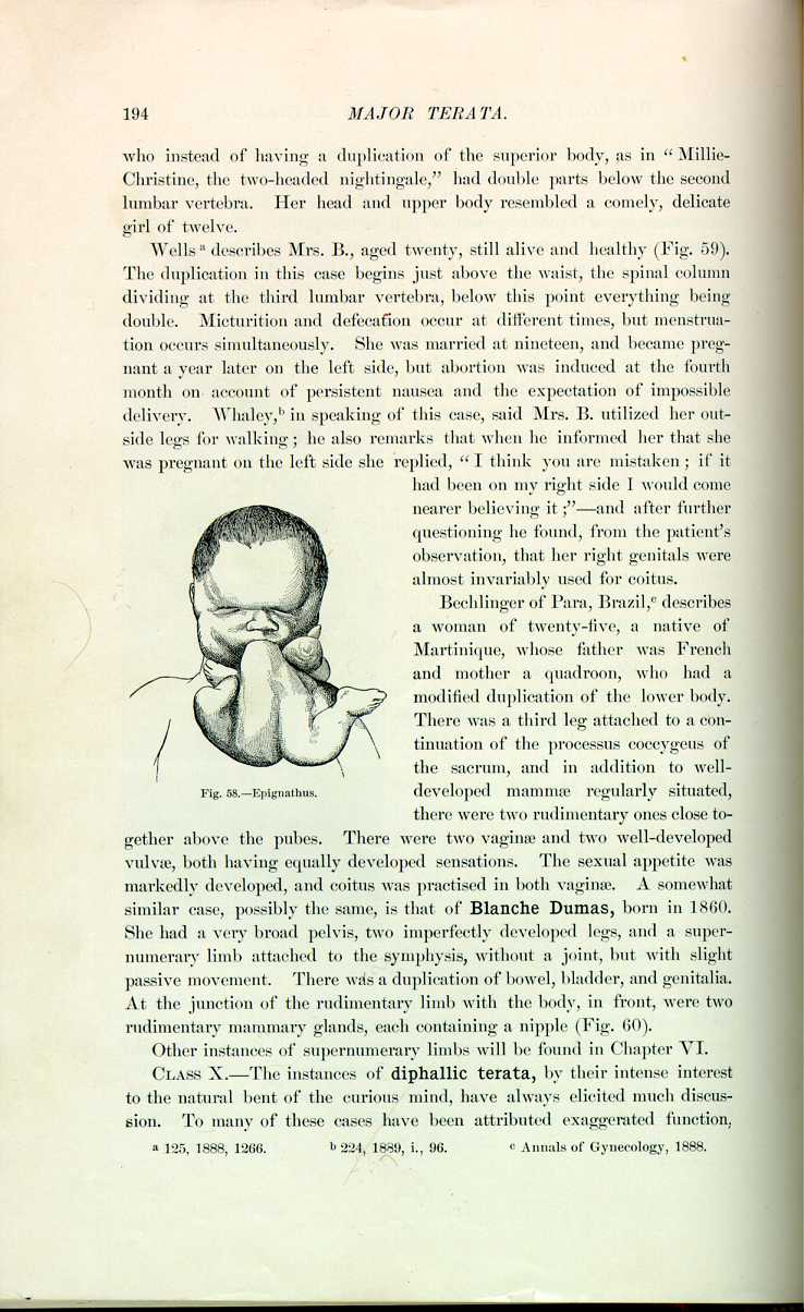
Fig. 58.—Epignathus.
[Description: Drawing of epignathus]
there were two rudimentary ones close together above the pubes. There were two vaginæ and two well-developed vulvæ, both having equally developed sensations. The sexual appetite was markedly developed, and coitus was practised in both vaginae. A somewhat similar case, possibly the same, is that of Blanche Dumas, born in 1860. She had a very broad pelvis, two imperfectly developed legs, and a supernumerary limb attached to the symphysis, without a joint, but with slight passive movement. There was a duplication of bowel, bladder, and genitalia. At the junction of the rudimentary limb with the body, in front, were two rudimentary mammary glands, each containing a nipple (Fig. 60).
Other instances of supernumerary limbs will be found in Chapter VI.
CLASS X.—The instances of diphallic terata, by their intense interest to the natural bent of the curious mind, have always elicited much discussion. To many of these cases have been attributed exaggerated function,
Schenck, Schurig, Bartholinus, Loder, and Ollsner report instances of diphallic terata; the latter case [5.57] was in a soldier of Charles VI., twenty-two years old, who applied to the surgeon for a bubonic affection, and who declared that he passed urine from the orifice of the left glans and also said that he was incapable of true coitus. Valentini mentions an instance in a boy of four, in which the two penises were superimposed. Bucchettoni [5.58] speaks of a man with

Fig, 59.—Dipygus (Wells).
[Description: Drawing of Mrs. B.]
two penises placed side by side. There was an anonymous case described [5.59] of a man of ninety-three with a penis which was for more than half its length divided into two distinct members, the right being somewhat larger than the left. From the middle of the penis up to the symphysis only the lower wall of the urethra was split. Jenisch [5.60] describes a diphallic infant, the offspring of a woman of twenty-five who had been married five years. Her first child was a well-formed female, and the second, the infant in question, cried much during the night, and several times vomited dark-green matter. In lieu of one penis there
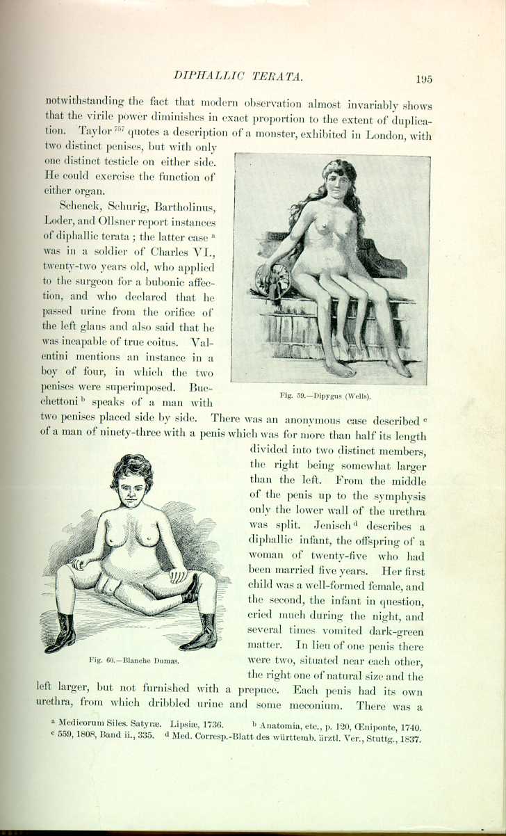
Fig. 60.—Blanche Dumas.
[Description: Drawing of Banche Dumas]
were two, situated near each other, the right one of natural size and the left larger, but not furnished with a prepuce. Each penis had its own urethra, from which dribbled urine and some meconium. There was a
Goré, reported by Velpeau, [5.61] has seen an infant of eight and one-half months with two penises and three lower extremities. The penises were 4 cm. apart and the scrotum divided, containing one testicle in each side. Each penis was provided with a urethra, urine being discharged from both simultaneously. In a similar case, spoken of by Geoffroy-Saint-Hilaire, the two organs were also separate, but urine and semen escaped sometimes from one, sometimes from both.
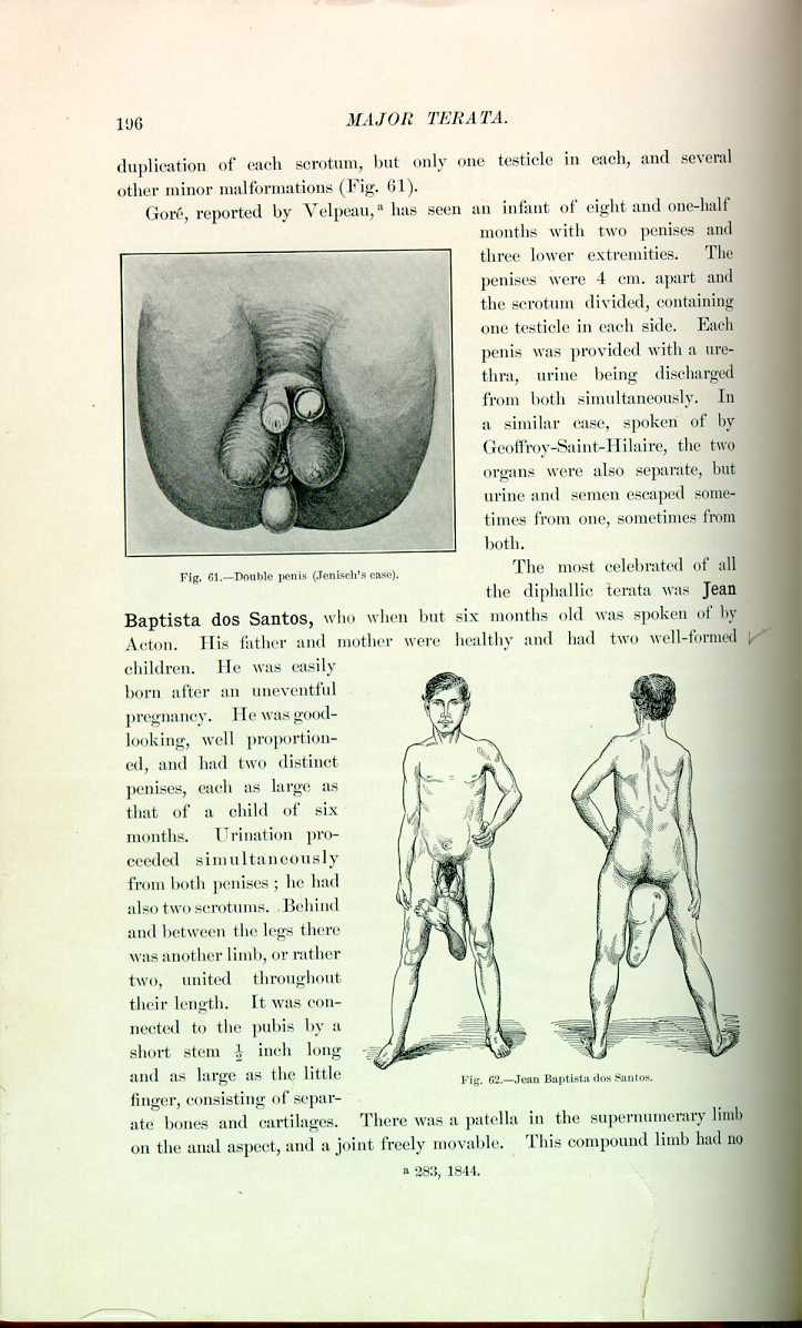
Fig. 61.—Double penis (Jenisch's case).
[Description: Drawing of a double penis]
The most celebrated of all the diphallic terata was Jean Baptista dos Santos, who when but six months old was spoken of by Acton. His father and mother were healthy and had two well-formed children. He was easily born after an uneventful pregnancy. He was good-looking, well proportioned, and had two distinct penises, each as large as that of a child of six months. Urination proceeded simultaneously from both penises; he had also two scrotums. Behind and between the legs there was another limb, or rather two, united throughout their length. It was connected to the pubis by a short stem 1/2 inch long and as large as the little

finger, consisting of separate bones and cartilages. There was a patella in the supernumerary limb on the anal aspect, and a joint freely movable. This compound limb had no
Van Buren and Keyes [5.65] describe a case in a man of forty-two, of good, healthy appearance. The two distinct penises of normal size were apparently well formed and were placed side by side, each attached at its root to the symphysis. Their covering of skin was common as far as the base of the glans; at this point they seemed distinct and perfect, but the meatus of the left was imperforate. The right meatus was normal, and through it most of the urine passed, though some always dribbled through an opening in the perineum at a point where the root of the scrotum should have been. On lifting the double-barreled penis this opening could be seen and was of sufficient size to admit the finger. On the right side of the aperture was an elongated and rounded prominence similar in outline to a labium majus. This prominence contained a testicle normal in shape and sensibility, but slightly undersized, and surrounded, as was evident from its mobility, by a tunica vaginalis. The left testicle lay on the tendon of the adductor longus in the left groin; it was not fully developed, but the patient had sexual desires, erections, and emissions. Both penises became erect simultaneously,
Sangalli [5.66] speaks of a man of thirty-five who had a supernumerary penis, furnished with a prepuce and capable of erection. At the apex of the glans opened a canal about 12 cm. long, through which escaped monthly a serous fluid. Smith [5.67] mentions a man who had two penises and two bladders, on one of which lithotomy was performed. According to Ballantyne, Taruffi, the scholarly observer of terata, mentions a child of forty-two months and height of 80 cm. who had two penises, each furnished with a urethra and well-formed scrotal sacs which were inserted in a fold of the groin. There were two testicles felt in the right scrotum and one in the left. Fecal evacuations escaped through two anal orifices. There is also another case mentioned similar to the foregoing in a man of forty; but here there was an osseous projection in the middle line behind the bladder. This patient said that erection was simultaneous in both penises, and that he had not married because of his chagrin over his deformity. Cole [5.68] speaks of a child with two well-developed male organs, one to the left and the other to the right of the median line, and about 1/4 or 1/2 inch apart at birth. The urethra bifurcated in the perineal region and sent a branch to each penis, and urine passed from each meatus. The scrotum was divided into three compartments by two raphes, and each compartment contained a testicle. The anus at birth was imperforate, but the child was successfully operated on, and at its sixtieth day weighed 17 pounds.
Lange [5.69] says that an infant was brought to Karg for relief of anal atresia when fourteen days old. It was found to possess duplicate penises, which communicated each to its distinct half of the bladder as defined by a median fold. The scrotum was divided into three portions by two raphes, and each lateral compartment contained a fully formed testicle. This child died because of its anal malformation, which we notice is a frequent associate of malformations or duplicity of the penis. There is an example in an infant described [5.70] in which there were two penises, each about 1/2 inch long, and a divided scrotal sac 21 inches long. Englisch [5.71] speaks of a German of forty who possessed a double penis of the bifid type.
Ballantyne and his associates define diphallic terata as individuals provided with two more or less well-formed and more or less separate penises, who may show also other malformations of the adjoining parts and organs (e. g., septate bladder), but who are not possessed of more than two lower limbs. This definition excludes, therefore, the cases in which in addition to a double penis there is a supernumerary lower extremity—such a case, for example, as that of Jean Baptista dos Santos, so frequently described by teratologists. It also excludes the more evident double terata, and, of course, the cases of
1. Diphallus, or duplication of the penis in an otherwise apparently single individual, is a very rare anomaly, records of only 20 cases having been found in a fairly exhaustive search through teratologic literature. As a distinct and well-authenticated type it has only quite recently been recognized by teratologists.
2. It does not of itself interfere with intrauterine or extrauterine life; but the associated anomalies (e. g., atresia ani) may be sources of danger. If not noticed at birth, it is not usually discovered till adult life, and even then the discovery is commonly accidental.
3. With regard to the functions of the pelvic viscera, urine may be passed by both penises, by one only, or by neither. In the last instance it finds exit by an aperture in the perineum. There is reason to believe that semen may be passed in the same way; but in most of the recorded cases there has been sterility, if not inability to perform the sexual act.
4. All the degrees of duplication have been met with, from a fissure of the glans penis to the presence of two distinct penises inserted at some distance from each other in the inguinal regions.
5. The two penises are usually somewhat defective as regards prepuce, urethra, etc.; they may lie side by side, or more rarely may be situated anteroposteriorly; they may be equal in size, or less commonly one is distinctly larger than the other; and one or both may be perforate or imperforate.
6. The scrotum may be normal or split; the testicles, commonly two in number, may be normal or atrophic, descended or undescended; the prostate may be normal or imperfectly developed, as may also the vasa deferentia and vesiculæ seminales.
7. The commonly associated defects are: More or less completely septate bladder, atresia ani, or more rarely double anus, double urethra, increased breadth of the bony pelvis with defect of the symphysis pubis, and possibly duplication of the lower end of the spine, and hernia of some of the abdominal contents into a perineal pouch. Much more rarely, duplication of the heart, lungs, stomach, and kidneys has been noted, and the lower limbs may be shorter than normal.
CLASS XI.—Cases of fetus in fetu, those strange instances in which one might almost say that a man may be pregnant with his brother or sister, or in
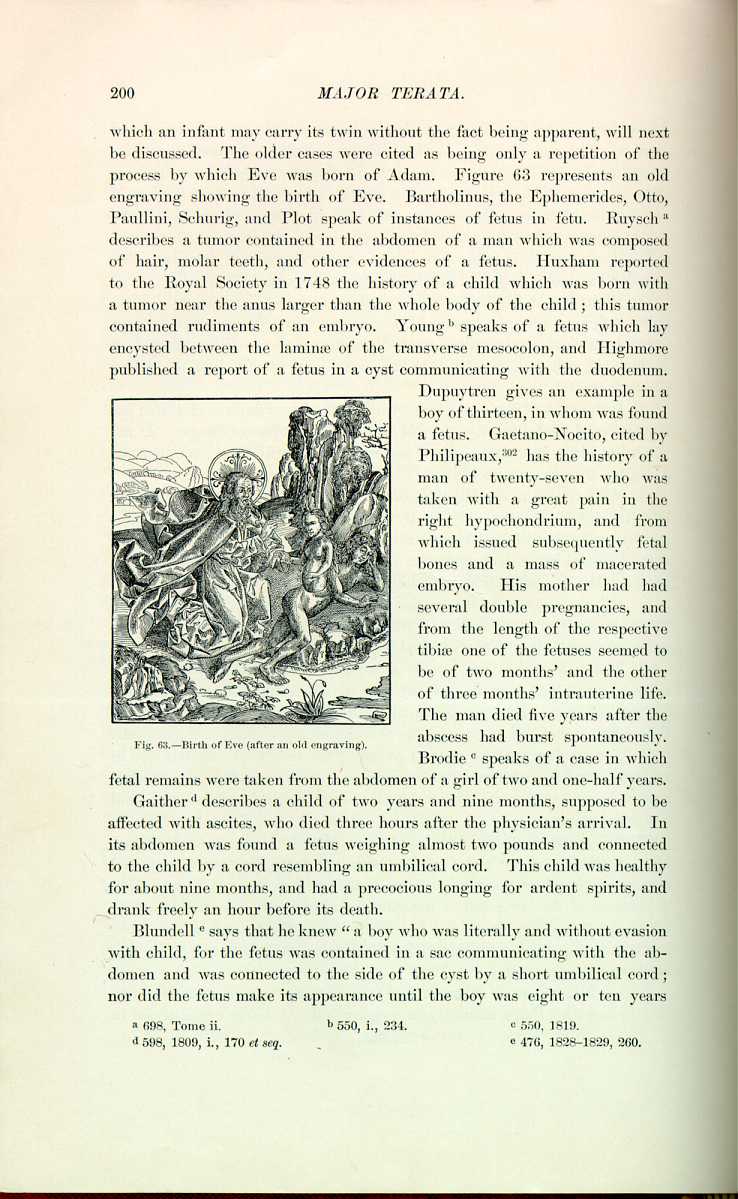
Fig. 63. -Birth of Eve (after an old engraving).
[Description: Engraving showing the birth of Eve]
Brodie [5.75] speaks of a case in which fetal remains were taken from the abdomen of a girl of two and one-half years. Gaither [5.76] describes a child of two years and nine months, supposed to be affected with ascites, who died three hours after the physician's arrival. In its abdomen was found a fetus weighing almost two pounds and connected to the child by a cord resembling an umbilical cord. This child was healthy for about nine months, and had a precocious longing for ardent spirits, and drank freely an hour before its death.
Blundell [5.77] says that he knew "a boy who was literally and without evasion with child, for the fetus was contained in a sac communicating with the abdomen and was connected to the side of the cyst by a short umbilical cord; nor did the fetus make its appearance until the boy was eight or ten years
At the Hôpital de la Charité in Paris, Velpeau startled an audience of 500 students and many physicians by saying that he expected to find a rudimentary fetus in a scrotal tumor placed in his hands for operation. His diagnosis proved correct, and brought him resounding praise, and all wondered as to his reasons for expecting a fetal tumor. It appears that he had read with care a report by Fatti [5.80] of an operation on the scrotum of a child which had increased in size as the child grew, and was found to contain the ribs, the vertebral column, the lower extremities as far as the knees, and the two orbits of a fetus; and also an account [5.81] of a similar operation performed by Wendt of Breslau on a Silesian boy of seven. The left testicle in this case was so swollen that it hung almost to the knee, and the fetal remains removed weighed seven ounces.
Sulikowski [5.82] relates an instance of congenital fetation in the umbilicus of a girl of fourteen, who recovered after the removal of the anomaly. Aretæos described to the members of the medical fraternity in Athens [5.83] the case of a woman of twenty-two, who bore two children after a seven months' pregnancy. One was very rudimentary and only 21 inches long, and the other had an enormous head resembling a case of hydrocephalus. On opening the head of the second fetus, another, three inches long, was found in the medulla oblongata, and in the cranial cavity with it were two additional fetuses, neither of which was perfectly formed.
Broca [5.84] speaks of a fetal cyst being passed in the urine of a man of sixty-one; the cyst contained remnants of hair, bone, and cartilage. Atlee [5.85] submits quite a remarkable case of congenital ventral gestation, the subject being a girl of six, who recovered after the discharge of the fetal mass from the abdomen. McIntyre [5.86] speaks of a child of eleven, playing about and feeling well, but whose abdomen progressively increased in size 1 1/2 inches each day. After ten days there was a large fluctuating mass on the right side; the abdomen was opened and the mass enucleated; it was found to contain a fetal mass weighing nearly five pounds, and in addition ten pounds of fluid were removed. The child made an early recovery. Rogers [5.87] mentions a fetus that
Modern literature is full of examples, and nearly every one of the foregoing instances could be paralleled from other sources. Rodriguez [5.90] is quoted as reporting that in July, 1891, several newspapers in the city of Mexico published, under the head of "A Man-mother,'' a wonderful story, accompanied by wood-cuts, of a young man from whose body a great surgeon had extracted a "perfectly developed fetus.'' One of these wood-cuts represented a tumor at the back of a man opened and containing a crying baby. In commenting upon this, after reviewing several similar cases of endocymian monsters that came under his observation in Mexico, Rodriguez tells what the case which had been so grossly exaggerated by the lay journals really was: An Indian boy, aged twenty-two, presented a tumor in the sacrococcygeal region measuring 53 cm. in circumference at the base, having a vertical diameter of 17 cm. and a transverse diameter of 13 cm. It had no pedicle and was fixed, showing unequal consistency. At birth this tumor was about the size of a pigeon's egg. A diagnosis of dermoid cyst was made and two operations were performed on the boy, death following the second. The skeleton showed interesting conditions; the rectum and pelvic organs were natural, and the contents of the cyst verified the diagnosis.
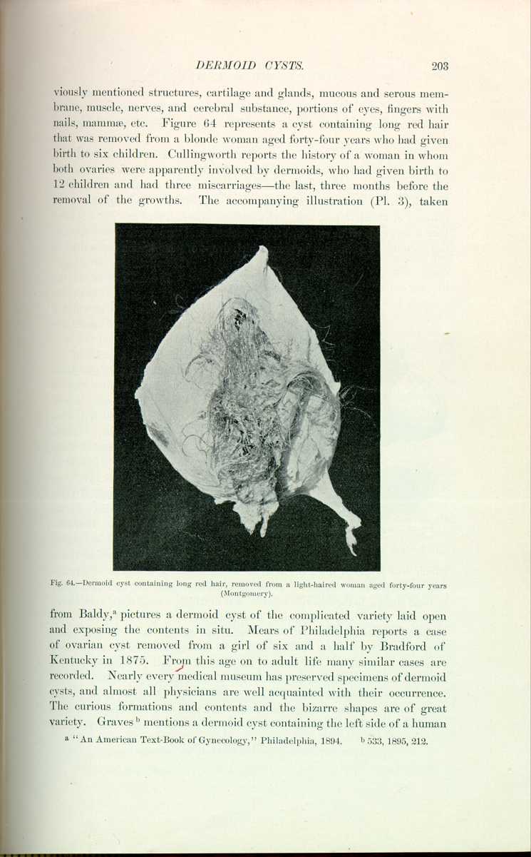
Quite similar to the cases of fetus in fetu are the instances of dermoid cysts. For many years they have been a mystery to physiologists, and their origin now is little more than hypothetic. At one time the fact of finding such a formation in the ovary of an unmarried woman was presumptive evidence that she was unchaste; but this idea was dissipated as soon as examples were reported in children, and to-day we have a well-defined difference between congenital and extrauterine pregnancy. Dermoid cysts of the ovary may consist only of a wall of connective tissue lined with epidermis and containing distinctly epidermic scales which, however, may be rolled up in firm masses of a more or less soapy consistency; this variety is called by Orth epidermoid cyst; or, according to Warren, a form of cyst made up of skin containing small and ill-defined papillæ, but rich in hair follicles and sebaceous glands. Even the erector pili muscle and the sudoriparous gland are often found. The hair is partly free and rolled up into thick balls or is still attached to the walls. A large mass of sebaceous material is also found in these cysts. Thomson reports a case of dermoid cyst of the bladder containing hair, which cyst he removed. It was a pedunculated growth, and it was undoubtedly vesical and not expelled from some ovarian source through the urinary passage, as sometimes occurs.
The simpler forms of the ordinary dermoid cysts contain bone and teeth. The complicated teratoma of this class may contain, in addition to the previously
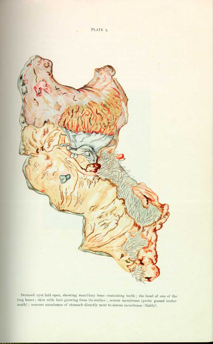
from Baldy, [5.91] pictures a dermoid cyst of the complicated variety laid open and exposing the contents in situ. Mears of Philadelphia reports a case of ovarian cyst removed from a girl of six and a half by Bradford of Kentucky in 1875. From this age on to adult life many similar cases are recorded. Nearly every medical museum has preserved specimens of dermoid cysts, and almost all physicians are well acquainted with their occurrence. The curious formations and contents and the bizarre shapes are of great variety. Graves [5.92] mentions a dermoid cyst containing the left side of a human
Weil reported the case of a man of twenty-two years who was born with what was supposed to he a spina bifida in the lower sacral region. According to Senn, the swelling never caused any pain or inconvenience until it inflamed, when it opened spontaneously and suppurated, discharging a large quantity of offensive pus, hair, and sebaceous material, thus proving it to have been a dermoid. The cyst was freely incised, and there were found numerous openings of sweat glands, from which drops of perspiration escaped when the patient was sweating.
Dermoid cysts of the thorax are rare. Bramann reported a case in which a dermoid cyst of small size was situated over the sternum at the junction of the manubrium with the gladiolus, and a similar cyst in the neck near the left cornu of the hyoid bone. Chitten removed a dermoid from the sternum of a female of thirty-nine, the cyst containing 11 ounces of atheromatous material. In the Museum of St. Bartholomew's Hospital in London
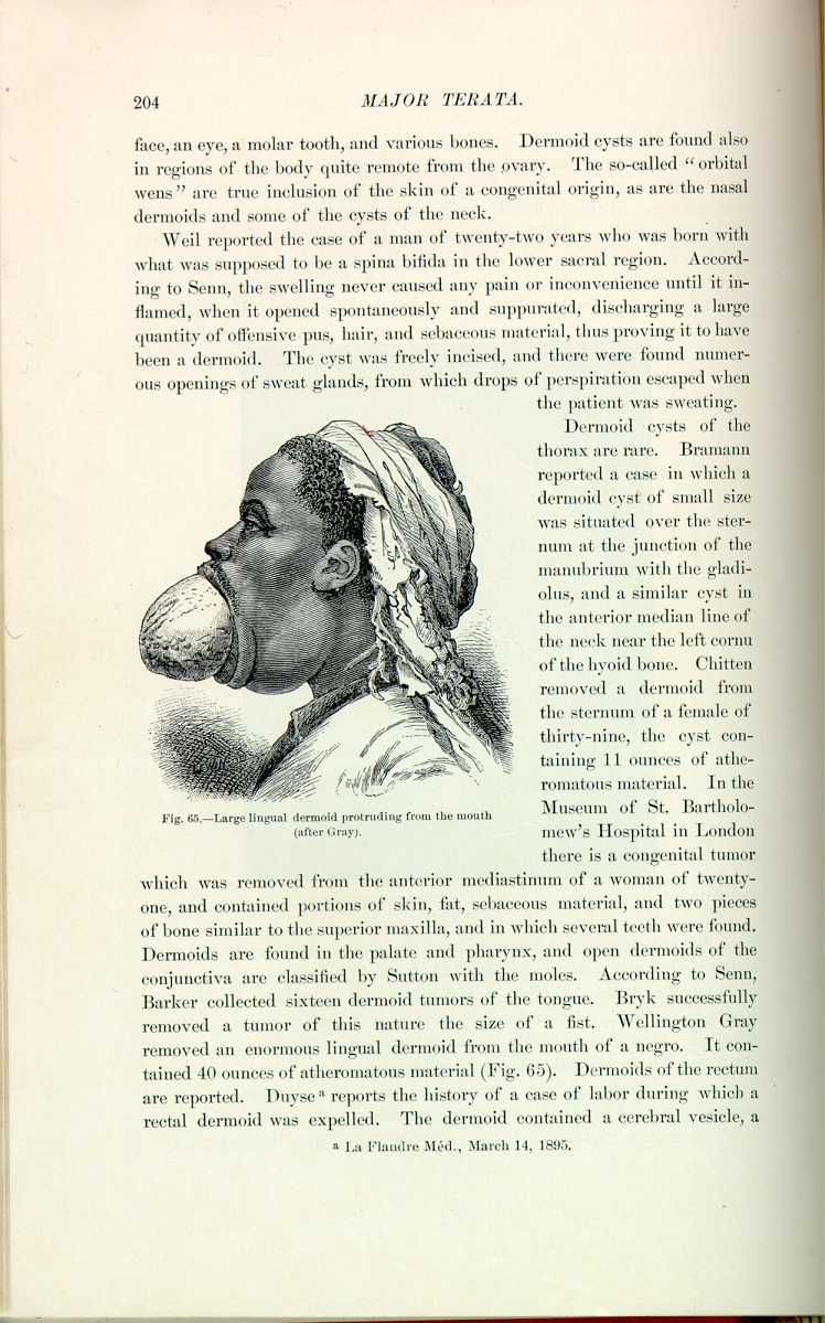
there is a congenital tumor which was removed from the anterior mediastinum of a woman of twenty one, and contained portions of skin, fat, sebaceous material, and two pieces of bone similar to the superior maxilla, and in which several teeth were found. Dermoids are found in the palate and pharynx, and open dermoids of the conjunctiva are classified by Sutton with the moles. According to Senn, Barker collected sixteen dermoid tumors of the tongue. Bryk successfully removed a tumor of this nature the size of a fist. Wellington Gray removed an enormous lingual dermoid from the mouth of a negro. It contained 40 ounces of atheromatous material (Fig. 65). Dermoids of the rectum are reported. Duyse [5.93] reports the history of a case of labor during which a rectal dermoid was expelled. The dermoid contained a cerebral vesicle, a
Ward [5.94] reports the successful removal of a dermoid cyst weighing 30 pounds from a woman of thirty-two, the mother of two children aged ten and twelve, respectively. The report is briefly as follows: "The patient has always been in good health until within the last year, during which time she has lost flesh and strength quite rapidly, and when brought to my hospital by her physician, Dr. James of Williamsburg, Kansas, was quite weak, although able to walk about the house. A tumor had been growing for a number of years, but its growth was so gradual that the patient had not considered her condition critical until quite recently. The tumor was diagnosed to be cystoma of the left ovary. Upon opening the sac with the trocar we were confronted by complications entirely unlooked for, and its use had to be abandoned entirely because the thick contents of the cyst would not flow freely, and the presence of sebaceous matter blocked the instrument. As much of the fluid as possible was removed, and the abdominal incision was enlarged to allow of the removal of the large tumor. An ovarian hematoma the size of a large orange was removed from the right side. We washed the intestines quite as one would wash linen, since some of the contents of the cyst had escaped into the abdominal cavity. The abdomen was closed without drainage, and the patient placed in bed without experiencing the least shock. Her recovery was rapid and uneventful. She returned to her home in four weeks after the operation.
"The unusual feature in this case was the nature of the contents of the sac. There was a large quantity of long straight hair growing from the cyst wall and an equal amount of loose hair in short pieces floating through the tumor-contents, a portion of which formed nuclei for what were called `moth-balls,' of which there were about 1 1/2 gallons. These balls, or marbles, varied from the size of moth-balls, as manufactured and sold by druggists, to that of small walnuts. They seemed to be composed of sebaceous matter, and were evidently formed around the short hairs by the motion of the fluid produced by walking or riding. There was some tissue resembling true skin attached to the inner wall of the sac.''
There are several cases of multiple dermoid cysts on record, and they may occur all over the body. Jamieson [5.95] reports a case in which there were 250, and in Maclaren's case there were 132. According to Crocker, Hebra and Rayer also each had a case. In a case of Sangster, reported by Politzer, although most of the dermoids, as usual, were like fibroma-nodules and therefore the color of normal skin, those over the mastoid processes and clavicle were lemon-yellow, and were generally thought to be xanthoma until they were excised, and Politzer found they were typical dermoid cysts with the usual contents of degenerated epithelium and hair.
Hermaphroditism.—Some writers claim that Adam was the first hermaphrodite and support this by Scriptural evidence. [5.96] We find in some of the ancient poets traces of an Egyptian legend in which the goddess of the moon was considered to be both male and female. From mythology we learn that Hermaphroditus was the son of Hermes, or Mercury, and Venus Aphrodite, and had the powers both of a father and mother. In speaking of the foregoing Ausonius writes, "Cujus erat facies in quâ paterque materque cognosci possint, nomen traxit ab illis.'' Ovid and Virgil both refer to legendary hermaphrodites, and the knowledge of their existence was prevalent in the olden times. The ancients considered the birth of hermaphrodites bad omens, and the Athenians threw them into the sea, the Romans, into the Tiber. Livy speaks of an hermaphrodite being put to death in Umbria, and another in Etruria. Cicero, Aristotle, Strabonius, and Pliny all speak concerning this subject. Martial [5.97] and Tertullian noticed this anomaly among the Romans. Aetius and Paulus ægineta speak of females in Egypt with prolonged clitorides which made them appear like hermaphrodites. Throughout the Middle Ages we frequently find accounts, naturally exaggerated, of double-sexed creatures. Harvey, Bartholinus, Paullini, Schenck, Wolff, Wrisberg, Zacchias, Marcellus Donatus, Haller, Hufeland, de Graff, and many others discuss hermaphroditism. Many classifications have been given, as, e. g., real and apparent; masculine, feminine, or neuter; horizontal and vertical; unilateral and bilateral, etc. The anomaly in most cases consists of a malformation of the external genitalia. A prolonged clitoris, prolapsed ovaries, grossness of figure, and hirsute appearance have been accountable for many supposed instances of hermaphrodites. On the other hand, a cleft scrotum, an ill-developed penis, perhaps hypospadias or epispadias, rotundity of the mammæ, and feminine contour have also provoked accounts of similar instances. Some cases have been proved by dissection to have been true hermaphrodites, portions or even entire genitalia of both sexes having been found.
Numerous accounts, many mythical, but always interesting, are given of these curious persons. They have been accredited with having performed the functions of both father and mother, notwithstanding the statements of some of the best authorities that they are always sterile. Observation has shown that the sexual appetite diminishes in proportion to the imperfections in the genitalia, and certainly many of these persons are sexually indifferent.
We give descriptions of a few of the most famous or interesting instances of hermaphroditism. Paré [5.98] speaks of a woman who, besides a vulva, from which she menstruated, had a penis, but without prepuce or signs of erectility. Haller alludes to several cases in which prolonged clitorides have been the cause of the anomaly. In commenting on this form of hermaphroditism Albucasiusus *[115] describes a necessary operation for the removal of the clitoris.
Beclard [5.102] showed Marie-Madeline Lefort, nineteen years of age, 1 1/2 meters in height. Her mammæ were well developed, her nipples erectile and surrounded by a brown areola, from which issued several hairs. Her feet were small, her pelvis large, and her thighs like those of a woman. Projecting from the vulva was a body looking like a penis 7 cm. long and slightly erectile at times; it was imperforate and had a mobile prepuce. She had a vulva with two well-shaped labia as shown by the accompanying illustration (Fig. 66). She menstruated slightly and had an opening at the root of the clitoris. The parotid region showed signs of a beard and she had

made (Fig. 68). There is no doubt but that this individual was Marie-Madeline Lefort in age.
Worbe [5.103] speaks of a person who was supposed to be feminine for twenty-two years. At the age of sixteen she loved a farmer's son, but the union was delayed for some reason, and three years later her grace faded and she became masculine in her looks and tastes. It was only after lengthy discussion, in which the court took part, that it was definitely settled that this person was a male.
Adelaide Préville, *[664] who was married as a female, and as such lived the last ten years of her life in France, was found on dissection at the Hôtel-Dieu
Fournier [5.105] speaks of an individual in Lisbon in 1807 who was in the highest degree graceful, the voice feminine, the mammæ well developed, The female genitalia were normal except the labia majora, which were rather diminutive. The thighs and the pelvis. were not so wide as those of a woman. There was some beard on the chin, but it was worn close. the male genitalia were of the size and appearance of
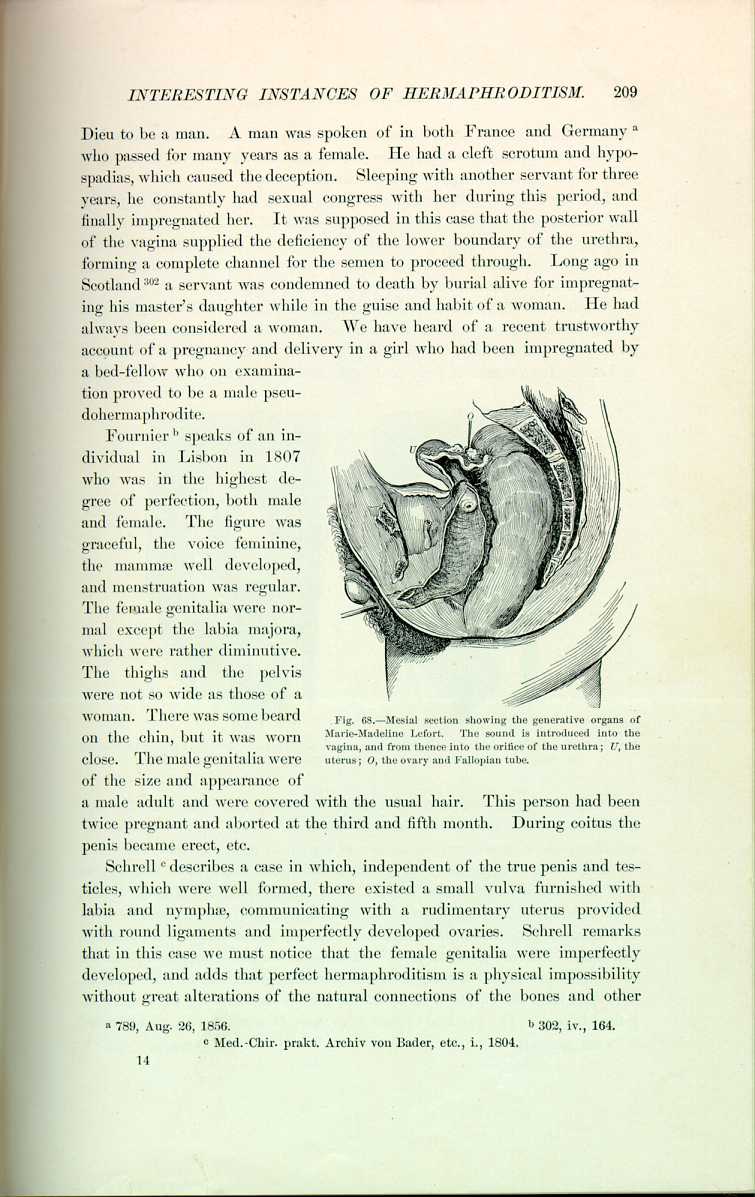
a male adult and were covered with the usual hair. This person had been twice pregnant and aborted at the third and fifth month. During coitus the penis became erect, etc.
Schrell [5.106] describes a case in which, independent of the true penis and testicles, which were well formed, there existed a small vulva furnished with labia and nymphæ, communicating with a rudimentary uterus provided with round ligaments and imperfectly developed ovaries. Schrell remarks that in this case we must notice that the female genitalia were imperfectly developed, and adds that perfect hermaphroditism is a physical impossibility without great alterations of the natural connections of the bones and other
In 1859 Hugier succeeded in restoring a vagina to a young girl of twenty who had an hypertrophied clitoris and no signs of a vagina. The accompanying illustrations show the conformation of the parts before operation with all the appearance of ill-developed male genitalia, and the appearance afterward with restitution of the vaginal opening (Fig. 69).
Virchow in 1872, Boddaert in 1875, and Marchand in 1883 report cases of duplication of the genitalia, and call their cases true hermaphrodites from an anatomic standpoint. There is a specimen in St. Bartholomew's Hospital in London from a man of forty-four, who died of cerebral hemorrhage. He
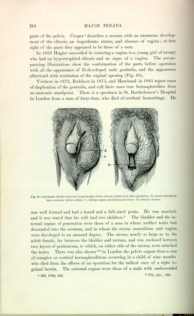
was well formed and had a beard and a full-sized penis. He was married, and it was stated that his wife had two children. [5.108] The bladder and the internal organs of generation were those of a man in whom neither testis had descended into the scrotum, and in whom the uterus masculinus and vagina were developed to an unusual degree. The uterus, nearly as large as in the adult female, lay between the bladder and rectum, and was enclosed between two layers of peritoneum, to which, on either side of the uterus, were attached the testes. There was also shown *[619] in London the pelvic organs from a case of complex or vertical hermaphroditism occurring in a child of nine months who died from the effects of an operation for the radical cure of a right inguinal hernia. The external organs were those of a male with undescended
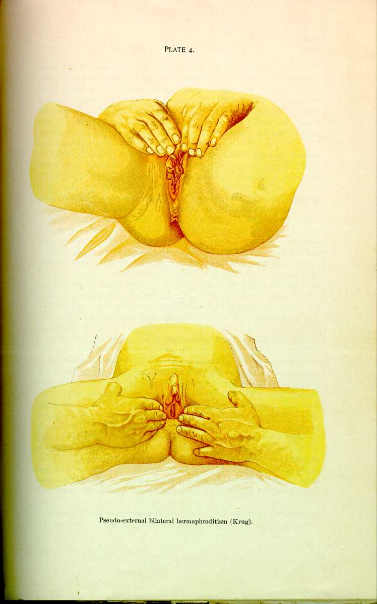
Barnes, Chalmers, Sippel, and Litten describe cases of spurious hermaphroditism due to elongation of the clitoris. In Litten's case [5.109] the clitoris was 3 1/2 inches long, and there was hydrocele of the processus vaginalis on both sides, making tumors in the labium on one side and the inguinal canal on the other, which had been diagnosed as testicles and again as ovaries. There was associate cystic ovarian disease. Plate 4 is taken from a case of false external bilateral hermaphroditism. Phillips [5.110] mentions four cases of spurious hermaphroditism in one family, and recently Pozzi [5.111] tells of a family of nine individuals in whom this anomaly was observed. The first was alive and had four children; the second was christened a female but was probably a male; the third, fourth, and fifth were normal but died young; the sixth daughter was choreic and feeble-minded, aged twenty-nine, and had one illegitimate child; the seventh, a boy, was healthy and married; the eighth was christened a female, but when seventeen was declared by the Faculty to be a male; the ninth was christened a female, but at eighteen the genitals were found to be those of a male, though the mammæ were well developed.
O'Neill [5.112] speaks of a case in which the clitoris was five inches long and one inch thick, having a groove in its inferior surface reaching down to an oblique opening in the perineum. The scrotum contained two hard bodies thought to be testicles, and the general appearance was that of hypospadias. Postmortem a complete set of female genitalia was found, although the ovaries were very small. The right round ligament was exceedingly thick and reached down to the bottom of the false scrotum, where it was firmly attached. The hard bodies proved to be on one side an irreducible omental hernia, probably congenital, and on the other a hardened mass having no glandular structure. The patient was an adult. As we have seen, there seems to be a law of evolution in hermaphroditism which prevents perfection. If one set of genitalia are extraordinarily developed, the other set are correspondingly atrophied. In the case of extreme development of the clitoris and approximation to the male type we must expect to find imperfectly developed uterus or ovaries. This would answer for one of the causes of sterility in these cases.
There is a type of hermaphroditism in which the sex cannot be definitely
The legal aspect of hermaphroditism has always been much discussed. Many interesting questions arise, and extraordinary complications naturally occur. In Rome a hermaphrodite could be a witness to a testament, the exclusive privilege of a man, and the sex was settled by the predominance. If the male aspect and traits together with the generative organs of man were most pronounced, then the individual could call himself a man. "Hermaphroditus an ad testamentum adhiberi possit qualitas sesus incalescentis ostendit.''
There is a peculiar case on record [5.114] in which the question of legal male inheritance was not settled until the individual had lived as a female for fifty-one years. This person was married when twenty-one, but finding coitus impossible, separated after ten years, and though dressing as a female had coitus with other women. She finally lived with her brother, with whom she eventually came to blows. She prosecuted him for assault, and the brother in return charged her with seducing his wife. Examination ensued, and at this ripe age she was declared to be a male.
The literature on hermaphroditism is so extensive that it is impossible to select a proper representation of the interesting cases in this limited space, and the reader is referred to the modern French works on this subject, in which the material is exhaustive and the discussion thoroughly scientific.
| CHAPTER V.
MAJOR TERATA. Anomalies and Curiosities of Medicine | ||