| The Origin and Nature of the Emotions: Miscellaneous Papers | ||
Histologic Changes in the Brain-cells in Relation to the Maintenance of Consciousness and to the Production of the Emotions, Muscular Activity, and Fever
We have studied the brain-cells in human cases of fever, and in animals after prolonged insomnia; after the injection of the toxins of gonococci, of streptococci, of staphylococci, and of colon, tetanus, diphtheria, and typhoid bacilli; and after the injection of foreign proteins, of indol and skatol, of leucin, and of peptones. We have studied the brains of animals which had been activated in varying degrees up to the point of complete exhaustion by running, by fighting, by rage and fear, by physical injury, and by the injection of strychnin (Figs. 2, 4, 5, and 37). We have studied the brains of salmon at the mouth of the Columbia River and at its headwater (Fig. 55); the brains of electric fish, the storage batteries of which had been partially discharged, and of those the batteries of which had been completely discharged; the brains of woodchucks in hibernation and after fighting; the brains of humans who had died from anemia resulting from hemorrhage, from acidosis, from eclampsia, from cancer and from other chronic diseases (Figs. 40 to 43, 56, 74, and 75). We have studied also the brains of animals after the excision of the adrenals, of the pancreas, and of the liver (Figs. 57 and 60).
In every instance the loss of vitality—that is, the loss of the normal power to convert potential into kinetic energy—was accompanied by physical changes in the brain-cells (Figs. 45 and 46). The converse was also true, that is, the brain-cells of animals with normal vital power showed no histologic changes. The changes in the brain-cells were
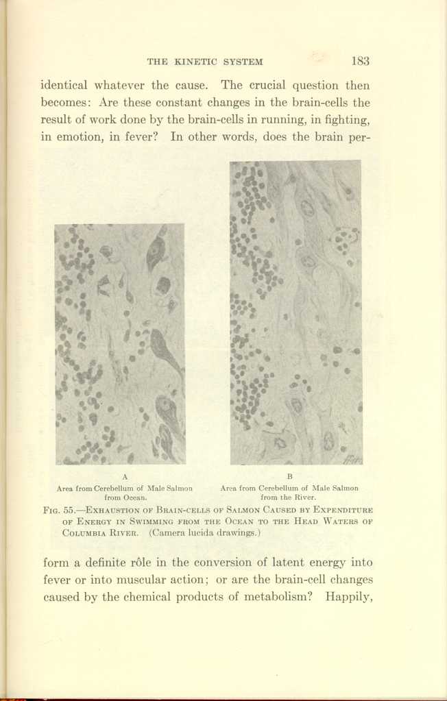
A: Area from Cerebellum of Male Salmon from Ocean.
B:Area from Cerebellum of Male Salmon from the River.
FIG. 55—EXHAUSTION OF BRAIN-CELLS OF SALMON CAUSED BY EXPENDITURE
OF ENERGY IN SWIMMING FROM THE OCEAN TO THE HEAD WATERS OF
COLUMBIA RIVER. (Camera lucida drawings.)
[Description: Black-and-white photographs showing cerebellum of salmon under
various conditions.]
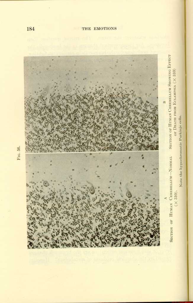
FIG. 56.
A: Section of Human Cerebellum—Normal (x310).
B: Section of Human Cerebellum Showing Effect of Death from Eclampsia
(x310).[a]
[Description: Black-and-white photographs showing human cerebellum under
various conditions.]
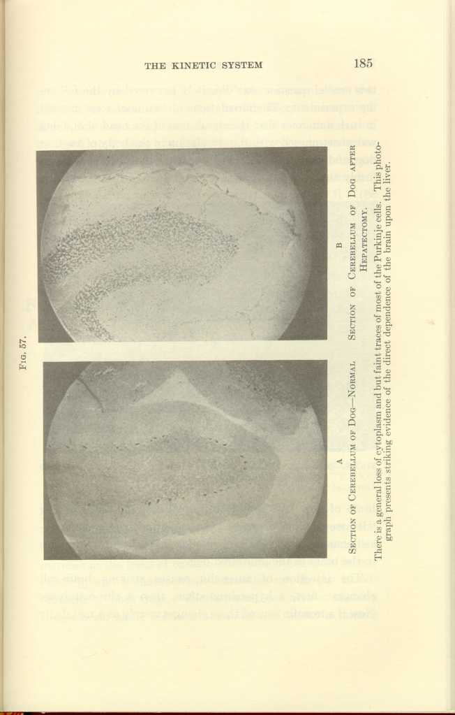
FIG. 57.
A: Section of Cerebellum of Dog—Normal
B: Section of Cerebellum of Dog after
Hepatectomy.[b]
[Description: Black-and-white photographs showing cerebellum of dog under
various conditions.]
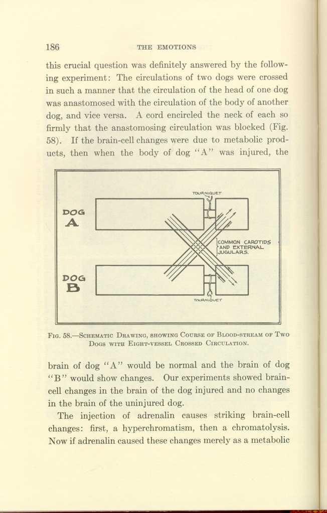
FIG. 58.—SCHEMATIC DRAWING, SHOWING COURSE OF BLOOD-STREAM OF TWO DOGS WITH EIGHT-VESSEL CROSSED CIRCULATION.
[Description: Black-and-white illustration: a diagram.]The injection of adrenalin causes striking brain-cell changes: first, a hyperchromatism, then a chromatolysis. Now if adrenalin caused these changes merely as a metabolic
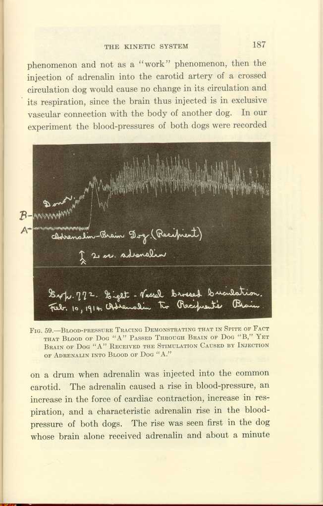
FIG. 59.—BLOOD-PRESSURE TRACING DEMONSTRATING THAT IN SPITE OF FACT THAT BLOOD OF DOG "A" PASSED THROUGH BRAIN OF DOG "B," YET BRAIN OF DOG "A" RECEIVED THE STIMULATION CAUSED BY INJECTION OF ADRENALIN INTO BLOOD OF DOG "A."
[Description: Black-and-white photograph of a chart.]1. Adrenalin alone causes hyperchromatism followed by chromatolysis, and in overdosage causes the destruction of some brain-cells.
2. When both adrenal glands are excised and no other factor is introduced, the Nissl substance progressively disappears from the brain-cells until death. This far-reaching point will be taken up later (Fig. 60).
Here our purpose is to discuss the cause of the brain-cell changes. We have seen that in crossed brain and body circulation trauma causes changes in the cells of the brain which is disconnected from the traumatized body by its circulation, but which is connected with the traumatized body by the nervous system. We have seen that adrenalin causes activation of the body connected with its brain by the nervous system, and histologic changes in the brain acted on directly by the adrenalin, but we found no notable brain-cell changes in the other brain through which the products of metabolism have circulated.
In the foregoing we find direct evidence that the products of metabolism are not the principal cause of the brain-cell changes. We shall now present evidence to show that for
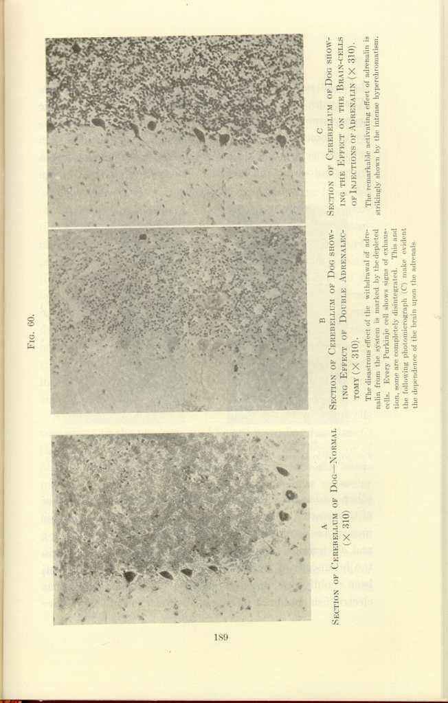
FIG. 60
A: Section of Cerebellum of Dog—Normal (x310).
B: Section of Cerebellum of Dog showing Effect of Double Adrenalectomy
(x310).[c]
C: Section of Cerebellum of Dog showing the Effect on the Brain-cells
of Injections of Adrenalin (x310).[d]
[Description: Black-and-white photographs showing cerebellum of dog under
various conditions.]
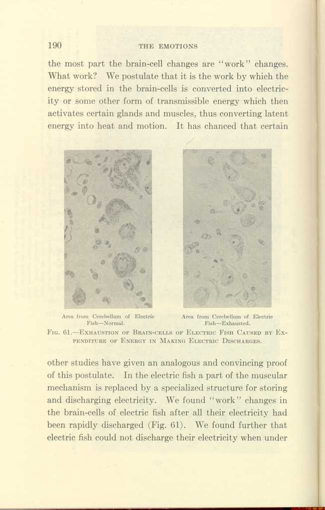
Area from Cerebellum of Electric Fish—Normal.
Area from Cerebellum of Electric Fish—Exhausted.
FIG. 61-EXHAUSTION OF BRAIN-CELLS OF ELECTRIC FISH CAUSED BY EXPENDITURE
OF ENERGY IN MAKING ELECTRIC DISCHARGES.
[Description: Black-and-white photographs showing cerebellum of fish under
normal and exhausted conditions.]
A further support to the postulate that the brain-cells contribute to the production of fever by sending impulses to the muscles is found in the effect of muscular exertion, or of other forms of motor stimulation, in the presence of a fever-producing infection. Under such circumstances muscular exertion causes additional fever, and causes also added but identical changes in the brain-cells. Thyroid extract and iodin have the same effect as muscular exertion and infection in the production of fever and the production of brain-cell changes. All this evidence is a strong argument in favor of the theory that certain constituents of the brain-cells
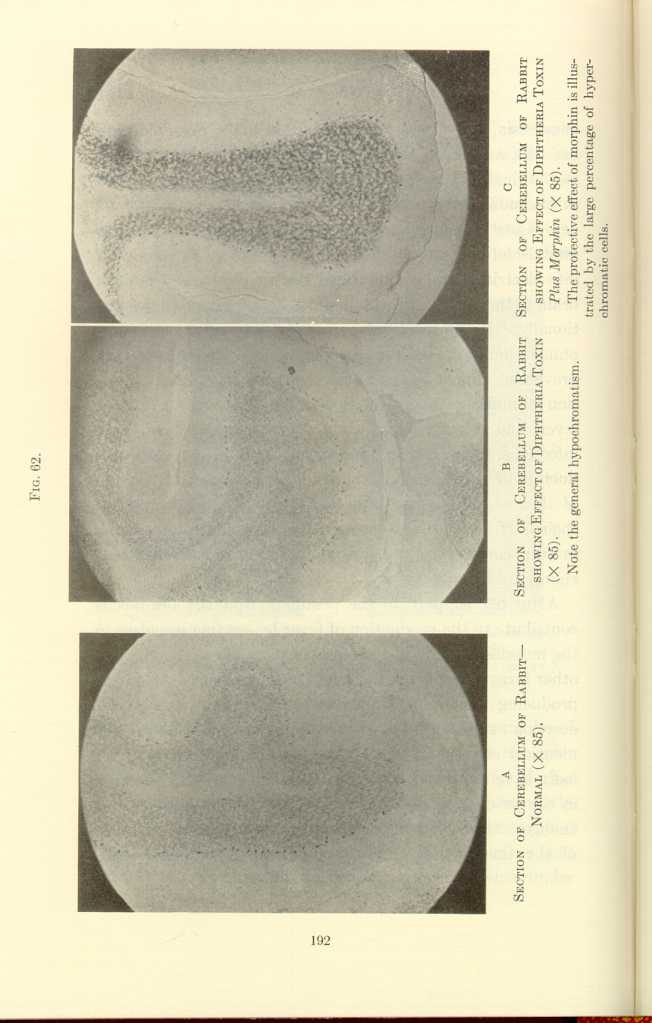
FIG. 62.
A: Section of Cerebellum of Rabbit—Normal (x85).
B: Section of Cerebellum of Rabbit showing Effect of Diptheria Toxin
(x85).[e]
C: Section of Cerebellum of Rabbit showing Effect of Diptheria Toxin
Plus Morphin (x85).[f]
[Description: Black-and-white photographs showing cerebellum of rabbit under
various conditions.]
That the stimulation of the brain-cells without gross activity of the skeletal muscles and without infection can produce heat is shown as follows:
(a) Fever is produced when animals are subjected to fear without any consequent exertion of the skeletal muscles.
(b) The temperature of the anxious friends of patients
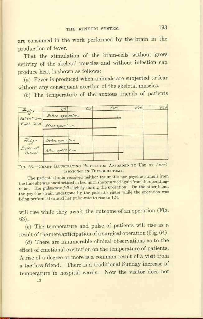
FIG. 63.-CHART ILLUSTRATING PROTECTION AFFORDED BY USE OF
Anoci-association IN THYROIDECTOMY.[g]
[Description: Black-and-white illustration: chart.]
(c) The temperature and pulse of patients will rise as a result of the mere anticipation of a surgical operation (Fig. 64).
(d) There are innumerable clinical observations as to the effect of emotional excitation on the temperature of patients. A rise of a degree or more is a common result of a visit from a tactless friend. There is a traditional Sunday increase of temperature in hospital wards. Now the visitor does not
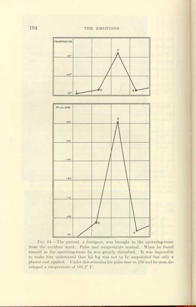
FIG. 64.[h]
[Description: Black-and-white illustration: two charts showing body temperature and pulse.]Is the contribution of the brain to the production of heat due to the conversion of latent energy directly into heat, or does the brain produce heat principally by converting its latent energy into electricity or some similar form of transmissible energy which, through nerve connections, stimulates other organs and tissues, which in turn convert their stores of latent energy into heat?
According to Starling, when the connection between the brain and the muscles of an animal is severed by curare, by anesthetics, by the division of the cord and nerves, then the heat-producing power of the animal so modified is on a level with that of cold-blooded animals. With cold the temperature falls, with heat it rises. Such an animal has no more control over the conversion of latent energy into heat than it has over the conversion of latent energy into motion.
Electric stimulation done over a period of time causes brain-cell changes, and electric stimulation of the muscles causes a rise in temperature.
| The Origin and Nature of the Emotions: Miscellaneous Papers | ||