| The Origin and Nature of the Emotions: Miscellaneous Papers | ||
THE RELATION BETWEEN THE PHYSICAL STATE OF THE BRAIN-CELLS AND BRAIN FUNCTIONS—EXPERIMENTAL AND CLINICAL[1]
The brain in all animals (including man) is but the clearing-house for reactions to environment, for animals are essentially motor or neuromotor mechanisms, composed of many parts, it is true, but integrated by the nervous system. Throughout the phylogenetic history of the race the stimuli of environment have driven this mechanism, whose seat of power—the battery—is the brain.
Since all normal life depends upon the response of the brain to the daily stimuli, we should expect in health, as well as in disease, to find modifications of the functions and the physical state of the component parts of this central battery—the brain-cells. Although we must believe, then, that every reaction to stimuli, however slight, produces a corresponding change in the brain-cells, yet there are certain normal, that is, non-diseased, conditions which produce especially striking changes. The cell changes due to the emotions, for example, are so similar, and in extreme conditions approach so closely to the changes produced by disease, that it is impossible to say where the normal ceases and the abnormal begins.
In view of the similarity of brain-cell changes it is not strange that in the clinic as well as in daily life, we are confronted constantly by outward manifestations which are so nearly identical that the true underlying cause of the condition
It is, therefore, the purpose of this paper to present the definite results of laboratory researches which show certain relations between alterations in brain functions and physical changes in the brain-cells.
Fear.—Our experiments have shown that the brain-cell changes due to fear may be divided into two stages: First, that of hyperchromatism—stimulation; second, that of hypochromatism—exhaustion (Figs. 5 and 13). Hyperchromatism was shown only in the presence of the activating stimuli or within a very short time after they had been received. This state gradually changed until the period of maximum exhaustion was reached—about six hours later. Then a process of reconstruction began and continued until the normal state was again reached.
Fatigue.—Fatigue from overexertion produced in the brain-cells like changes to those produced by fear, these changes being proportional to the amount of exertion (Fig. 4). In the extreme stage of exhaustion from this cause we found that the total quantity of Nissl substance was enormously reduced. When the exertion was too greatly prolonged, it took weeks or months for the cells to be restored to their normal condition. We have proved, therefore, that in exhaustion resulting from emotion or from physical work
Hemorrhage.—The loss of blood from any cause, if sufficient to reduce the blood-pressure, will occasion a change in the brain-cells, provided that the period of hypotension lasts for more than five minutes. This time limit is a safeguard against permanent injury from the temporary hypotension which causes one to faint. If the hemorrhage be long continued and the blood-pressure be low, there will be a permanent loss of some of the brain-cells. This explains why an individual who has suffered from a prolonged hemorrhage will never again be restored to his original powers.
Drugs.—According to their effect upon the brain-cells, drugs may be divided into three classes: First, those that stimulate the brain-cells to increased activity, as strychnin (Fig. 37); second, those that chemically destroy the brain-cells, as alcohol and iodoform (Figs. 38 and 39); third, those that suspend the functions of the cells without damaging them, as nitrous oxid, ether, morphin. Our experiments have shown that the brain-cell changes induced by drugs of the first class are precisely the same as the cycle of changes produced by the emotions and by physical activity. We have found that strychnin, according to the dosage, causes convulsions ending in exhaustion and death; excitation followed by lassitude; stimulation without notable after-results; or
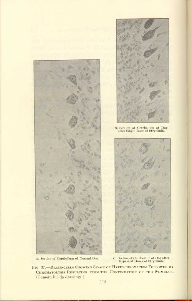
A, Section of Cerebellum of Normal Dog.
B, Section of Cerebellum of Dog after Single Dose of Strychnin.
C, Section of Cerebellum of Dog after Repeated Doses of Strychnin.
FIG. 37.—BRAIN-CELLS SHOWING STAGE OF HYPERCHROMATISM FOLLOWED BY
CHROMATOLYSIS RESULTING FROM THE CONTINUATION OF THE STIMULUS.
(Camera lucida drawings.)
[Description: Black-and-white photographs showing cerebellum of god under
various conditions.]
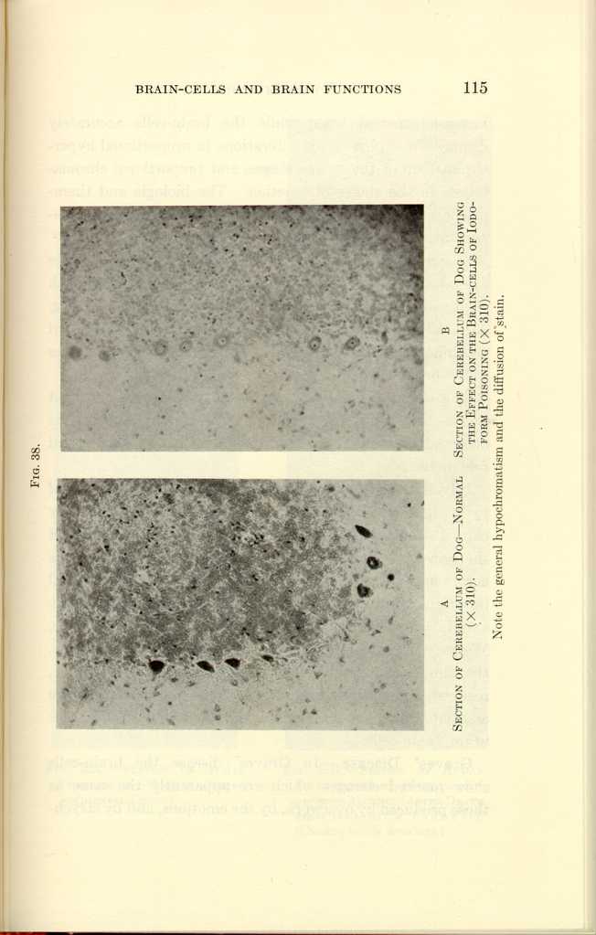
FIG. 38.
A: Section of Cerebellum of Dog—Normal (x310).
B: Section of Cerebellum of Dog Showing the Effect of the Brain-cells
of Iodoform Poisoning (x310).[a]
[Description: Black-and-white photographs of cerebellum of dog under various
conditions.]
In our experiments, alcohol in large and repeated dosage caused marked morphologic changes in the brain-cells which went as far even as the destruction of some of the cells (Fig. 39). Ether, on the other hand, even after five hours of administration, produced no observable destructive changes in the brain-cells.
The effect of iodoform was peculiarly interesting, as it was the only drug that produced a rise of temperature. Its observed effect upon the brain-cells was that of wide-spread destruction.
Infections.—In every observation regarding the effect of pyogenic infections on dogs and on man we found that they caused definite and demonstrable lesions in certain cells of the nervous system, the most marked changes being in the cortex and the cerebellum (Fig. 40). For example, in fatal infections resulting from bowel obstruction, in peritonitis, and in osteomyelitis, the real lesion is in the brain-cells. We may, therefore, reasonably conclude that the lassitude, the diminished mental power, the excitability, irritability, restlessness, delirium, and unconsciousness which may be associated with acute infections, are due to physical changes in the brain-cells.
Graves' Disease.—In Graves' disease the brain-cells show marked changes which are apparently the same as those produced by overwork, by the emotions, and by strychnin.
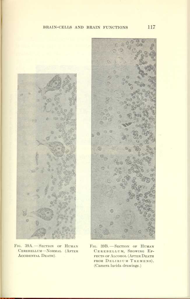
FIG. 39A.—SECTION OF HUMAN CEREBELLUM—NORMAL (AFTER ACCIDENTAL
DEATH).
FIG. 39B.—SECTION OF HUMAN CEREBELLUM, SHOWING EFFECTS OF ALCOHOL
(AFTER DEATH FROM DELIRIUM TREMENS). (Camera lucida drawings.)
[Description: Black-and-white photographs showing human cerebellum under
various conditions.]
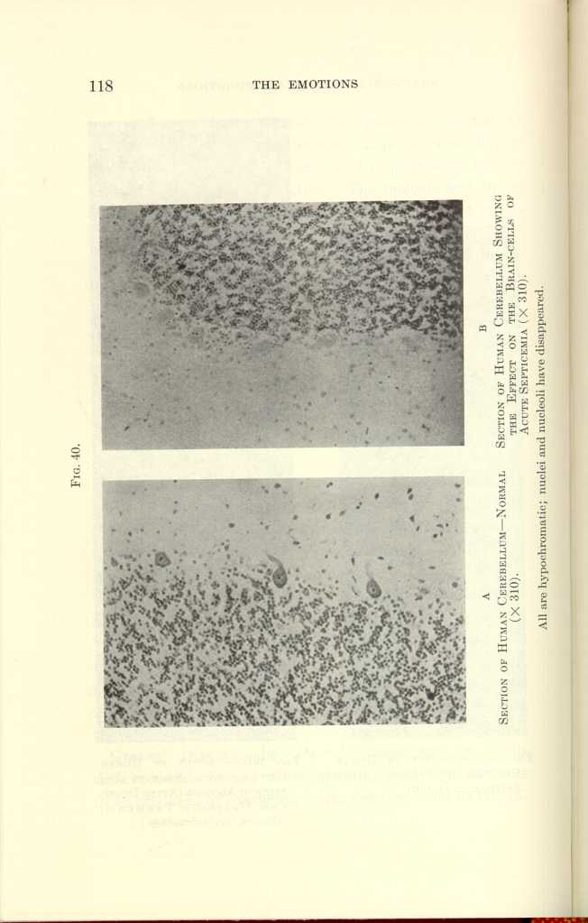
FIG. 40.
A: Section of Human Cerebellum—Normal (x310).
B: Section of Human Cerebellum Showing the Effect on the Brain-cells of
Acute Septicemia (x310).[b]
[Description: Black-and-white photographs of human cerebellum under various
conditions.]
Insomnia.—The brains of rabbits which had been kept awake for one hundred hours showed precisely the same changes as those shown in physical fatigue, strychnin poisoning, and exhaustion from emotional stimulation. Eight hours of continuous sleep restored all the cells except those that had been completely exhausted. This will explain the permanent ill effect of long-continued insomnia; that is, long-continued insomnia permanently destroys a part of the brain-cells just as do too great physical exertion, certain drugs, emotional strain, exophthalmic goiter, and hemorrhage. We found, however, that if, instead of natural sleep, the rabbits were placed for the same number of hours under nitrous oxid anesthesia, not only did the brain-cells recover from the physical deterioration, but that 90 per cent. of them became hyperchromatic. This gives us a possible clue to the actual chemical effect of sleep. For since nitrous oxid owes its anesthetic effect to its influence upon oxidation, we may infer that sleep also retards the oxidation of the cell contents. If this be true, then it is probable that inhalation anesthetics exert their peculiar influence upon that portion of the brain through which sleep itself is produced. If nitrous oxid anesthesia and sleep are chemically identical, then we have a further clue to one of the primary mechanisms of life itself; and as a practical corollary one might be able to produce artificial sleep which would closely resemble
In the case of the rabbit in which nitrous oxid was substituted for sleep, the appearance of the brain-cells resembled that in but one other group experimentally examined—the brain-cells of hibernating woodchucks.
Insanity.—Our researches have shown that in the course of a fatal disease and in fatal exhaustion, however produced, death does not ensue until there is marked disorganization of the brain tissue. In the progress of disease or exhaustion one may see in different patients every outward manifestation of mental deterioration, manifestations which, in a person who does not show any other sign of physical disease, mark him as insane. Take, for example, the progressive mental state of a brilliant scholar suffering from typhoid fever. On the first day of the gradual onset of the disease he would notice that his mental power was below its maximum efficiency; on the second he would notice a further deterioration, and so the mental effect of his disease would progress until he would find it impossible to express a thought or to make a deduction. No one can be philanthropic with jaundice; no one suffering from Graves' disease can be generous; no mental process is possible in the course of the acute infectious diseases. Just prior to death from any cause every one is in a mental state which, if it could be continued, would cause that individual to be judged insane. If the delirium that occurs in the course of certain diseases should be continued, the patient would be judged insane. In severe cases of Graves' disease the patient is insane.
Our experiments have proved conclusively that whether we call a person fatigued or diseased, the brain-cells undergo physical deterioration, accompanied by loss of mental power (Figs. 40 to 43). Even to the minutest detail we can show a direct relationship between the physical state of the brain-cells and the mental power of the individual, that is, the physical power of a person goes pari passu with his mental power. Indeed, it is impossible to conceive how any mental action, however subtle, can occur without a corresponding change in the brain-cells. It is possible now to measure only the evidences of the effects on the brain-cells of gross and violent mental activity. At some future time it will doubtless be possible so to refine the technic of brain-cell examinations
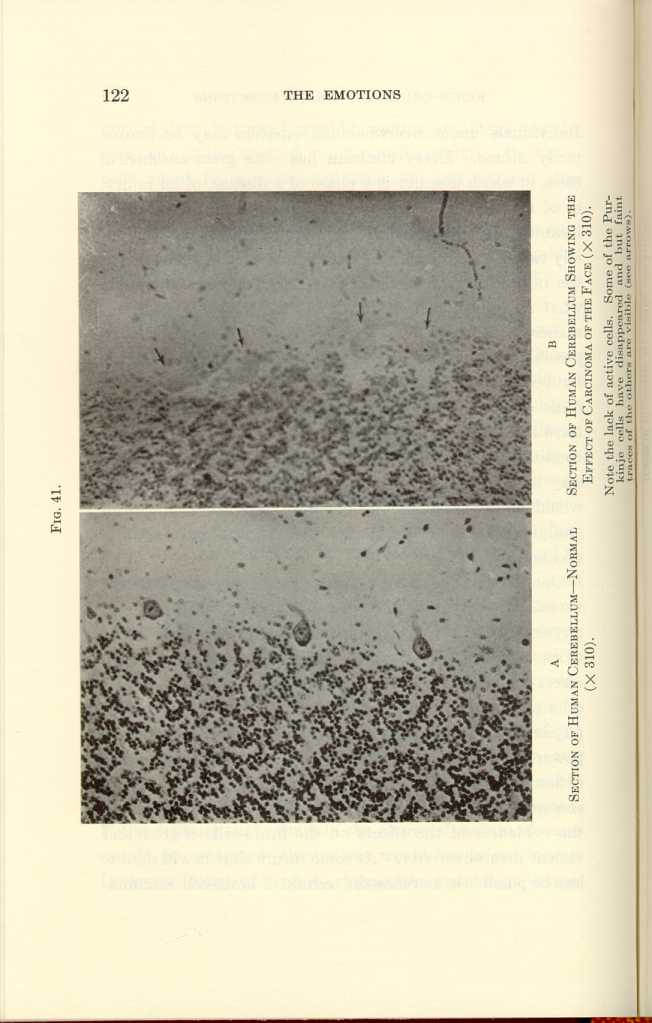
FIG. 41.
A: Section of Human Cerebellum—Normal (x310).
B: Section of Human Cerebellum Showing the Effect of Carcinoma of the
Face (x310).[c]
[Description: Black-and-white photographs showing human cerebellum under various
conditions.]
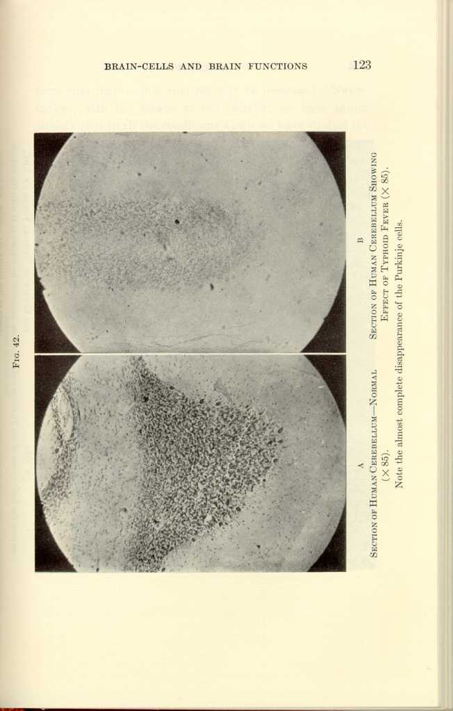
FIG. 42.
A: Section of Human Cerebellum—Normal (x85).
B: Section of Human Cerebellum Showing Effect of Typhoid Fever
(x85).[d]
[Description: Black-and-white photographs showing human cerebellum under
various conditions.]
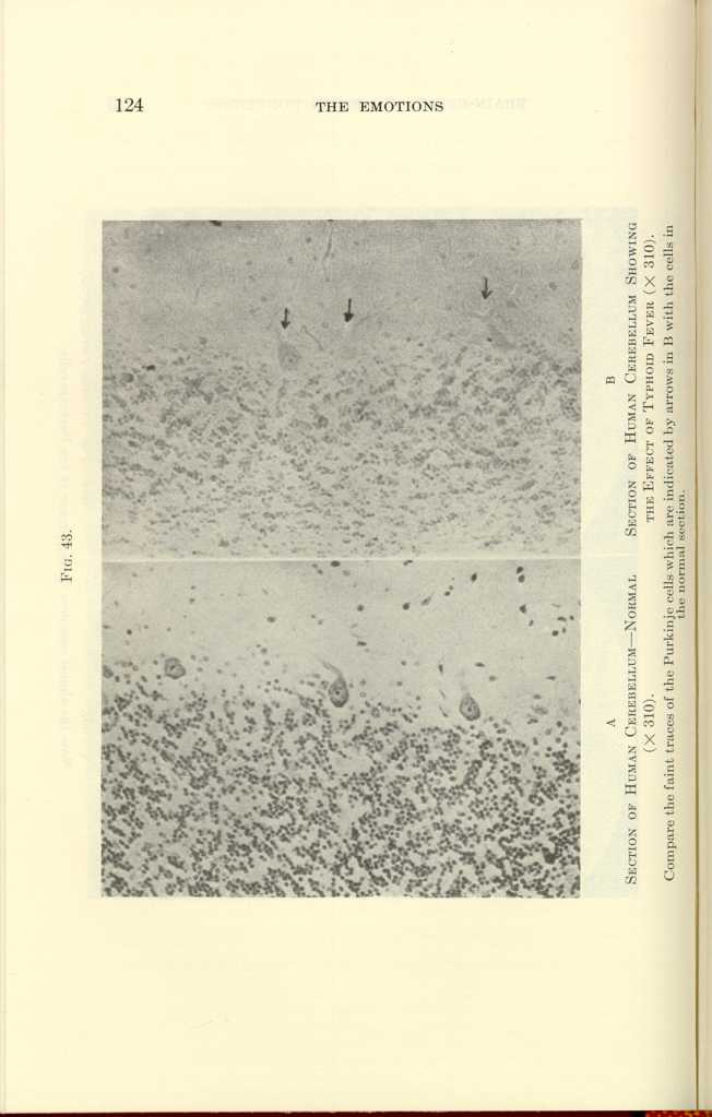
FIG. 43.
A: Section of Human Cerebellum—Normal (x310).
B: Section of Human Cerebellum Showing the Effect of Typhoid Fever
(x310).[e]
[Description: Black-and-white photographs showing human cerebellum under
various conditions.]
| The Origin and Nature of the Emotions: Miscellaneous Papers | ||